
Eyelid Agenesis
Eyelid agenesis is a hereditary disease, most commonly seen in cats, and it may affect several kittens from the same litter. Affected animals are born with an incomplete eyelid margin. The disorder is generally bilateral and symmetrical, and the defect involves all eyelid layers, most commonly in the middle portion of the upper eyelid. In dogs, the lateral portion of the lower eyelid is most commonly affected. The size of the defect may vary.
This genetic anomaly is often accompanied by other ocular abnormalities, with persistent pupillary membranes being the most common associated problem in cats. In dogs, eyelid agenesis may occasionally be accompanied by microphthalmia, absence of the lacrimal glands, persistent pupillary membranes, cataract, retinal dysplasia, and optic nerve coloboma. In some dogs, a complete eyelid margin may be present but without developed Meibomian glands.
Eyelid agenesis most commonly results in trichiasis (contact between hair and the ocular surface), which can lead to corneal changes, vascularization, pigmentary keratitis, corneal sequestrum, and ulceration. If an animal cannot completely close the eye with the eyelids, the protective function of the eyelids is lost. The eye is then exposed to adverse environmental influences and drying. Such animals are predisposed to ocular trauma and keratitis.
Treatment
In some mild cases, frequent application of lubricants to the ocular surface and permanent removal of the hair follicles producing hairs that irritate the eye are sufficient. If the patient has pronounced clinical problems, is unable to protect the cornea with the eyelids, or develops recurrent corneal ulcers despite preventive measures, the ophthalmologist may recommend surgical reconstruction of the eyelids.
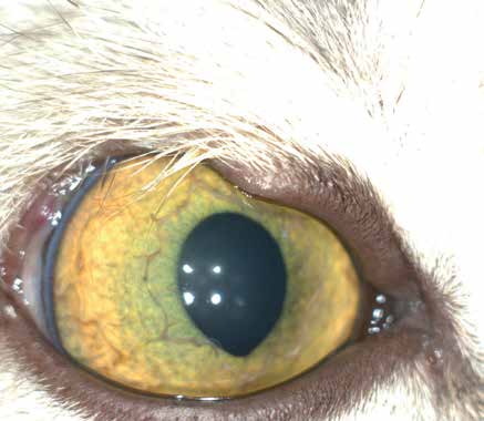
Agenesis of one half of the upper eyelid in a cat. The eyelid margin is absent, allowing hairs from the upper eyelid to contact the cornea (trichiasis).
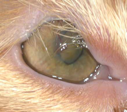
Agenesis of two thirds of the upper eyelid in a cat. The eyelid margin is absent, allowing hairs from the upper eyelid to contact the cornea (trichiasis). The cornea is chronically irritated by the hairs, with neovascularization, pigmentation, and corneal scarring. Ocular discharge is present as a result of irritation.
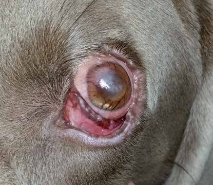
Agenesis of one half of the upper eyelid in a dog (medial region). The eyelid margin is absent, allowing hairs from the upper eyelid to contact the cornea (trichiasis). The cornea is chronically irritated by the hairs, with neovascularization and chronic central fibrosis. As a result of the agenesis, the orbicularis oculi muscle is underdeveloped, leading to reduced eyelid motility and chronic keratitis.
Adapted from the Atlas of Ocular Diseases in Dogs and Cats by Dr. Siniša D. Grozdanić and colleagues. © Animal Eye Consultants of Iowa. The text and clinical images on this page are original content from the authors' own clinical practice and research, and may not be reproduced, distributed, or repurposed without permission.
Think your pet may have eyelid agenesis?
Early evaluation gives us the most treatment options and the best chance of preserving vision.