
Extraocular Myositis (EOM)
Extraocular myositis, periocular myositis, and masticatory muscle myositis are inflammatory diseases of muscle that may affect one muscle group or several groups. The disease is most commonly autoimmune in nature. Extraocular myositis may also be caused by the protozoa Neospora caninum or Toxoplasma gondii. It is most commonly diagnosed in German Shepherd Dogs, Labrador Retrievers, Golden Retrievers, Shih Tzus, Lhasa Apsos, and other dog breeds.
Changes may be bilateral or unilateral and are sometimes accompanied by abnormalities of general health, including apathy, anorexia, and increased body temperature. Dogs may develop acute, painful swelling of affected muscles, exophthalmos, and frequent protrusion of the third eyelid. Periocular swelling may be very pronounced and may occasionally resemble an abscess. Pain or restriction during opening of the jaw is very common. Without aggressive treatment, muscle fibrosis and atrophy may develop, together with compressive optic nerve damage and vision loss.
Treatment
Diagnosis can be made on the basis of clinical and ultrasonographic examination. Immunosuppressive medications are used for treatment.
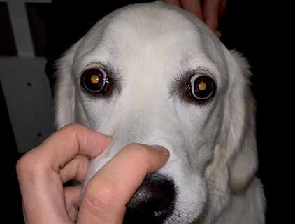
Extraocular myositis in a young dog.
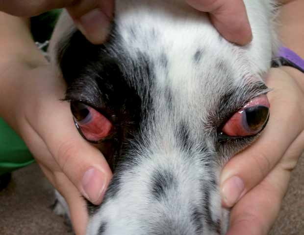
Extraocular myositis with exophthalmos, protrusion of the third eyelid, and conjunctival hyperemia.
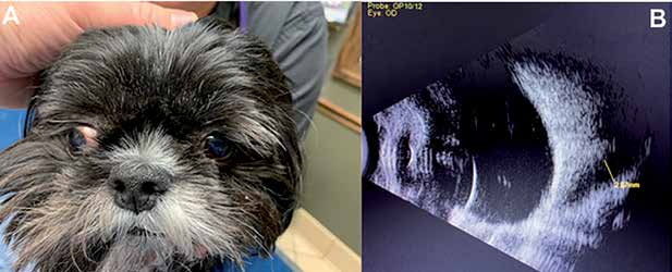
A) Unilateral extraocular myositis - the right eye is exophthalmic and has divergent strabismus. B) Ultrasonographic image of the same dog showing swelling of an extraocular muscle (black hypoechoic region marked by the yellow line).
Adapted from the Atlas of Ocular Diseases in Dogs and Cats by Dr. Siniša D. Grozdanić and colleagues. © Animal Eye Consultants of Iowa. The text and clinical images on this page are original content from the authors' own clinical practice and research, and may not be reproduced, distributed, or repurposed without permission.
Think your pet may have extraocular myositis (eom)?
Early evaluation gives us the most treatment options and the best chance of preserving vision.