
Eversion of the Third Eyelid Cartilage
The third eyelid is supported by T-shaped cartilage. The cartilage can occasionally bend, twist, or evert, deforming the third eyelid, particularly its free margin. This condition is most commonly seen in young dogs of large breeds. In German Shorthaired Pointers, it is hereditary. Prolapse of the third eyelid gland may develop secondary to this condition.
Treatment
Surgical correction is the most reliable treatment. The deformed cartilage is straightened using thermocautery or the bent margins of the cartilage are excised.
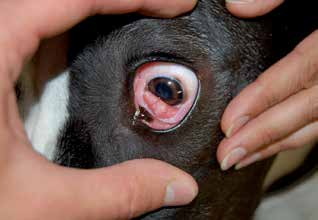
Eversion of the third eyelid cartilage.
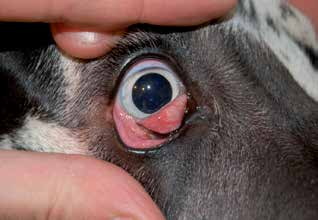
Eversion of the third eyelid cartilage.
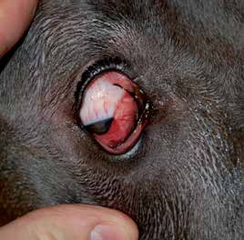
Eversion of the third eyelid cartilage.
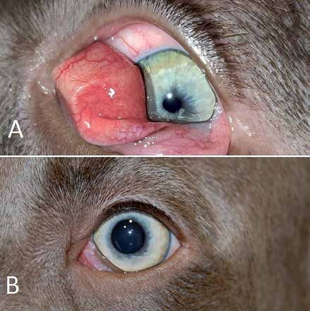
A) Eversion of the third eyelid cartilage and prolapse of the third eyelid gland before surgery. B) The same dog after cartilage correction with thermocautery and repositioning of the prolapsed gland.
Adapted from the Atlas of Ocular Diseases in Dogs and Cats by Dr. Siniša D. Grozdanić and colleagues. © Animal Eye Consultants of Iowa. The text and clinical images on this page are original content from the authors' own clinical practice and research, and may not be reproduced, distributed, or repurposed without permission.
Think your pet may have eversion of the third eyelid cartilage?
Early evaluation gives us the most treatment options and the best chance of preserving vision.