
Ectopic Cilium (Eyelash)
An ectopic cilium is an additional hair or group of hairs that emerges through the palpebral conjunctiva and is directed toward the ocular surface. This abnormality may be observed in young dogs, is more common in the upper eyelid, and is usually unilateral. A breed predisposition exists; breeds in which ectopic cilia are commonly encountered include the Boxer, English and French Bulldog, Shih Tzu, and Golden Retriever.
An ectopic cilium may cause keratitis and persistent corneal ulcers accompanied by excessive tearing and conjunctival swelling. Ectopic cilia are not always easy to identify, particularly in uncooperative dogs or those with marked conjunctival swelling. They may also be difficult to see in dogs with light-colored coats, because the cilium may also be light in color. If an ectopic cilium is suspected, the palpebral conjunctiva should be examined carefully under magnification using a slit lamp or operating microscope.
Treatment
The ectopic cilium should be removed and the hair follicle destroyed. Manual epilation without permanent removal of the follicle provides only a temporary solution. The follicle can be eliminated by cryotherapy, electroepilation, or surgery. Keratitis and/or corneal ulceration caused by the ectopic cilium must also be treated.
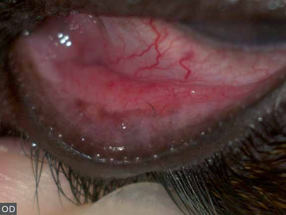
Several ectopic cilia protruding from the conjunctiva of the lower eyelid. They are easily visible because the hairs are black against a lighter background.
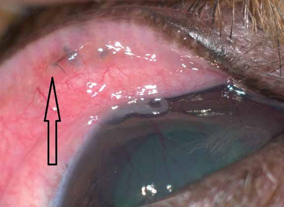
Group of ectopic cilia at multiple locations on the conjunctiva of the upper eyelid. Dark localized islands represent cilia located sub-conjunctivally, and a smaller number of ectopic cilia protrude and irritate the cornea. Corneal vascularization and pathological ocular discharge are present.
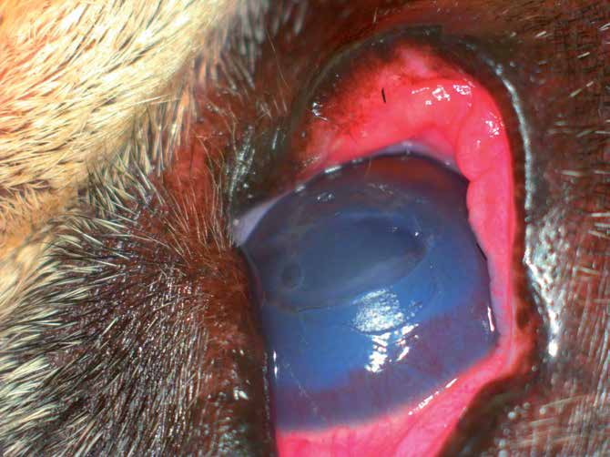
A single ectopic cilium on the conjunctiva of the upper eyelid directed toward the corneal surface and causing severe irritation. Conjunctival swelling and hyperemia, corneal vascularization, and a large corneal ulcer are present.
Adapted from the Atlas of Ocular Diseases in Dogs and Cats by Dr. Siniša D. Grozdanić and colleagues. © Animal Eye Consultants of Iowa. The text and clinical images on this page are original content from the authors' own clinical practice and research, and may not be reproduced, distributed, or repurposed without permission.
Think your pet may have ectopic cilium (eyelash)?
Early evaluation gives us the most treatment options and the best chance of preserving vision.