
Dry Eye Disease (Keratoconjunctivitis Sicca)
The tear film has an aqueous component secreted by the lacrimal glands, as well as lipid and mucin components secreted by eyelid glands and conjunctival goblet cells. Dry eye disease is a progressive inflammatory condition of the cornea and conjunctiva caused by tear deficiency. The deficiency may be qualitative, resulting from inadequate lipid and mucin components, or quantitative, resulting from reduced secretion by the lacrimal glands.
The causes of dry eye disease are numerous. The condition may develop secondary to autoimmune and allergic disease, inflammatory disorders, certain infectious diseases such as canine distemper, endocrine disorders including hypothyroidism, hyperadrenocorticism (Cushing's disease), and diabetes mellitus, nasolacrimal duct obstruction, neurologic disorders, toxins or certain medications such as sulfonamides, and removal of the third eyelid gland.
Affected animals develop conjunctivitis with pronounced conjunctival hyperemia and swelling and keratitis that is often pigmentary and/or ulcerative. Corneal vascularization is present, and corneal scarring can substantially affect visual quality. Without tears as a protective component, the eye is susceptible to bacterial infection, and mucopurulent ocular discharge is therefore common. The animal experiences discomfort or pain and blepharospasm is present. Changes are most commonly bilateral but differ in severity between the eyes. Dogs are affected more frequently than cats; when dry eye disease occurs in cats, the clinical signs are generally less pronounced. Certain dog breeds are predisposed, and dry eye disease is diagnosed more often in middle-aged and older dogs.
Treatment
Tear quantity is measured with the Schirmer tear test, while tear-film quality can be assessed using tear-film breakup time. Treatment includes topical lacrimostimulants and artificial tears. The primary cause is addressed whenever possible, allergies are controlled, and medications responsible for the condition are discontinued. If medical treatment does not provide satisfactory results, parotid duct transposition may be performed. In this procedure, the salivary duct is surgically moved from the oral cavity to the lower eyelid so that saliva can lubricate the ocular surface.
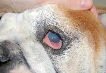
Dry eye disease in an English Bulldog. Corneal scarring resulting from chronic inflammation and keratitis with vascularization are present. The conjunctiva is inflamed, the third eyelid gland is prolapsed, and ocular discharge is retained on the eyelid skin. The Schirmer tear test result is 0 mm/min.
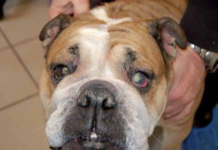
Dry eye disease in an English Bulldog. This dog has no third eyelid glands; they were removed one year earlier. The dog also has atopy and combined ectropion and entropion, all of which contributed to development of dry eye disease. A substantial amount of purulent discharge is present in the left eye. The Schirmer tear test result is 0 mm/min.
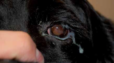
Dry eye disease in a dog. The Schirmer tear test value is 7 mm/min. Conjunctival hyperemia, a substantial amount of discharge, and reduced corneal transparency caused by chronic inflammation are visible.
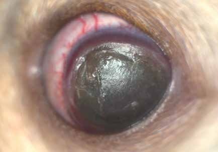
Very advanced dry eye disease in a dog that cannot blink. The cornea is insensitive and a large, deep stromal ulcer is present.
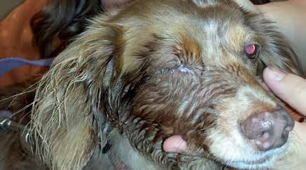
Parotid duct transposition in a patient with an absolutely dry eye resulted in excessive salivation and moist dermatitis of the facial skin. Surgical reduction of the parotid duct lumen was performed in this case and substantially reduced wetting of the face.
Adapted from the Atlas of Ocular Diseases in Dogs and Cats by Dr. Siniša D. Grozdanić and colleagues. © Animal Eye Consultants of Iowa. The text and clinical images on this page are original content from the authors' own clinical practice and research, and may not be reproduced, distributed, or repurposed without permission.
Think your pet may have dry eye disease (keratoconjunctivitis sicca)?
Early evaluation gives us the most treatment options and the best chance of preserving vision.