
Drug-Induced Retinal Injury
The source states that multiple medications and compounds can produce retinal changes. Some may cause reversible tapetal color changes, such as ethambutol, while others may cause serious irreversible injury and vision loss, and lists dithizone, ivermectin, moxidectin, and isoxazoline products.
In cats, the source identifies irreversible retinal degeneration secondary to enrofloxacin as a major concern, particularly at high doses, while noting that blindness has also been reported below 5 mg/kg/day. Mydriasis, loss of pupillary and palpebral reflexes as described in the source, and sudden vision loss may develop after days to weeks of treatment. Cats in which enrofloxacin is used for a shorter duration or stopped promptly at the first sign of mydriasis or visual difficulty are described as having a better chance of visual recovery. Retinal degeneration may continue to progress even after treatment is discontinued. Changes can involve all retinal layers, particularly photoreceptors and retinal pigment epithelium. Fundic findings include granularity and tapetal hyperreflectivity, golden-yellow or copper-colored spots, loss and clumping of nontapetal pigment, progressive attenuation and loss of retinal vessels, and optic nerve atrophy.
In dogs, the source states that ivermectin, particularly in overdose or after prolonged use, can cause retinal degeneration and most commonly reversible blindness. The fundus may appear macroscopically normal, or optic disc swelling, retinal edema, and focal retinal detachment may be present. The appearance may resemble multifocal retinal dysplasia. Visual impairment ranges from partial deficits to complete blindness depending on exposure. The source also describes oral isoxazoline flea and tick products, including afoxolaner, fluralaner, lotilaner, and sarolaner, as a cause of retinal toxicity in dogs and states that the changes are often reversible after discontinuation, although permanent retinal lesions may remain in some dogs.
Treatment
Diagnosis is based on ophthalmic examination, ERG findings, and history. No specific treatment is described. If a dog is receiving ivermectin, administration is discontinued and intravenous lipid therapy may be attempted in acute intoxication according to the source.
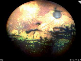
Retinal degeneration in a cat two months after enrofloxacin treatment. Hyperreflective fundic areas, reduced pigmentation and pigment clumping in the nontapetal region, and optic nerve atrophy are visible.
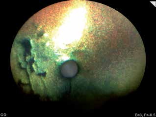
Retinal changes in a cat after enrofloxacin treatment. Retinal vessels are no longer visible and the entire tapetal region is hyperreflective. This cat is blind.
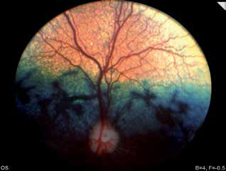
Retinopathy in a dog after ivermectin treatment. Fundic appearance two months after administration.
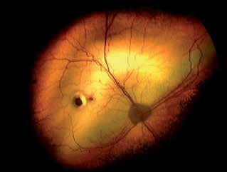
Ivermectin-associated retinopathy in a Border Collie. Horseshoe-shaped tapetal hyperreflectivity and a scar with pigment accumulation are visible.
Related in-depth guide
Retinal Disorders
Our full guide, including treatment and surgical options
Adapted from the Atlas of Ocular Diseases in Dogs and Cats by Dr. Siniša D. Grozdanić and colleagues. © Animal Eye Consultants of Iowa. The text and clinical images on this page are original content from the authors' own clinical practice and research, and may not be reproduced, distributed, or repurposed without permission.
Think your pet may have drug-induced retinal injury?
Early evaluation gives us the most treatment options and the best chance of preserving vision.