
Dacryocystitis
Dacryocystitis is inflammation of the lacrimal sac and nasolacrimal duct. It may develop as a consequence of primary bacterial infection but most commonly occurs secondary to stenosis or obliteration of the lacrimal duct. The duct may narrow or become obstructed because of a foreign body such as a grass awn, facial bone fracture, neoplasia of the sinuses or facial bones compressing the nasolacrimal duct, or periodontal disease of the premolars.
Epiphora, most commonly unilateral, dominates the clinical presentation. Chronic mucopurulent ocular discharge may subsequently develop. Swelling may occur in the region of the lacrimal sac if a cyst or abscess forms, and this swelling may be painful on palpation.
Treatment
Obstruction of the nasolacrimal ducts can be assessed using the relatively unreliable Jones fluorescein test: fluorescein is placed in the eye and, if the nasolacrimal duct is patent, should appear at the ipsilateral nostril within 10-15 minutes. Diagnosis can also be made by irrigation of the nasolacrimal duct through the lacrimal punctum. Reflux of mucopurulent material indicates obstruction; if mucopurulent material appears at the opposite opening, inflammation of the nasolacrimal duct is suspected.
Radiography, MRI, and CT may also be used, and are more successful if contrast medium is introduced into the nasolacrimal duct. After diagnosis, the cause of the obstruction is addressed. More forceful irrigation can often remove an obstruction such as a foreign body or accumulated exudate. Catheterization or specialized forceps may sometimes help. Surgical access to the duct may occasionally be necessary to remove the cause of obstruction. Topical and systemic antibiotics and anti-inflammatory medications are administered after these procedures. If patency cannot be restored, a fistula between the lacrimal sac and nasal cavity must be created to provide an alternative route for tear drainage.
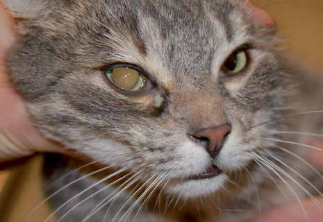
Dacryocystitis in a cat.
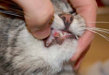
Damaged canine tooth that caused dacryocystitis in the cat shown in the previous figure.
Adapted from the Atlas of Ocular Diseases in Dogs and Cats by Dr. Siniša D. Grozdanić and colleagues. © Animal Eye Consultants of Iowa. The text and clinical images on this page are original content from the authors' own clinical practice and research, and may not be reproduced, distributed, or repurposed without permission.
Think your pet may have dacryocystitis?
Early evaluation gives us the most treatment options and the best chance of preserving vision.