
Congenital Glaucoma
Glaucoma represents a combination of progressive ocular pathologic processes that impair vision and produce characteristic changes in the optic nerve, and that are associated with elevated intraocular pressure. In veterinary medicine, glaucoma is one of the most common causes of vision loss according to the source. Glaucoma involves reduced aqueous-humor outflow, increased intraocular pressure, reduced blood supply to the optic nerve head, structural changes in the optic nerve head, and neuroinflammation with abnormalities of retinal ganglion cells.
Because of scleral elasticity, high intraocular pressure causes enlargement of the globe, or buphthalmos, particularly in young animals such as puppies and kittens and in adults with chronic glaucoma. The cornea becomes thicker and cloudy because of stromal edema. The optic nerve undergoes anoxia, axonal death occurs, and gliosis develops at the optic nerve head. Glaucoma is classified as congenital, primary, or secondary.
Congenital glaucoma is inherited and present at birth. Patients show signs of glaucoma shortly after birth, within a few weeks or months. Both eyes are affected, although changes need not be symmetric. Clinical signs include increased intraocular pressure, mydriasis, corneal edema with possible Haab's striae, secondary ulcerative keratitis, episcleral vascular congestion, buphthalmos, lens subluxation, secondary uveitis, cortical cataract, vitreous degeneration, retinal degeneration, and optic nerve head atrophy.
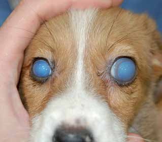
Congenital glaucoma in a dog. Buphthalmos is visible in the left eye and corneal edema is visible in both eyes.
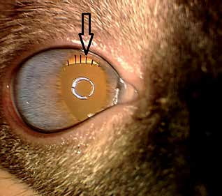
Congenital glaucoma in a cat with buphthalmos and elongation of the ciliary processes, indicated by the black arrow.
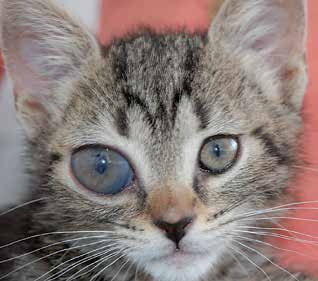
Congenital glaucoma in a cat. Buphthalmos of the right eye and corneal edema are visible.
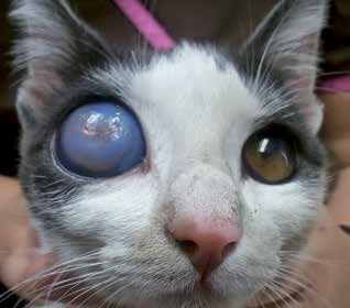
Congenital glaucoma in a cat with pronounced buphthalmos, corneal edema, and corneal erosion secondary to lagophthalmos.
Treatment
Diagnosis is established by ophthalmic examination, with tonometry being particularly important. Gonioscopy and high-frequency ultrasonography can help determine whether glaucoma is primary or secondary. Medical treatment consists of antiglaucoma eye drops intended to reduce aqueous-humor production or increase its outflow. For a visual eye in which intraocular pressure cannot be adequately controlled medically, the source describes transscleral laser cyclophotocoagulation combined with placement of an anterior chamber shunt as the surgical procedure of choice. If the eye is blind, has permanent morphologic changes, and has no realistic potential for recovery of vision, enucleation is recommended because of uncontrolled glaucoma and associated pain.
Related in-depth guide
Glaucoma
Our full guide, including treatment and surgical options
Adapted from the Atlas of Ocular Diseases in Dogs and Cats by Dr. Siniša D. Grozdanić and colleagues. © Animal Eye Consultants of Iowa. The text and clinical images on this page are original content from the authors' own clinical practice and research, and may not be reproduced, distributed, or repurposed without permission.
Think your pet may have congenital glaucoma?
Early evaluation gives us the most treatment options and the best chance of preserving vision.