
Clinical Examination of the Eye
The eye is a mirror of health and should never be evaluated as an isolated organ; the general health status of the pet must always be taken into account. Ocular changes are often a consequence of life-threatening pathological conditions, whether the eye is the primary site of disease or merely reflects systemic disease. Likewise, a dog that is not systemically endangered but has visual problems and/or ocular irritation is not a happy dog. Such a pet may spend more time sleeping, may be less interested and less active, and routine activities may become unpleasant or potentially dangerous. Owners are often unaware of the degree of discomfort their pet had until they visit an ophthalmologist and the problem is resolved. Only then do they recognize the change in their pet, noticing that it is more playful and appears "rejuvenated." For this reason, a detailed and professional general clinical examination of the dog or cat, together with a detailed and professional ophthalmic examination, is necessary for accurate diagnosis and selection of therapy.
A detailed ocular examination should include all or some of the following steps:
- Careful history taking
- Ophthalmic examination
- Specialized diagnostic testing
Every examination begins with the history. Information regarding breed, age, sex, medical history, current or recent therapy, travel or changes in place of residence, and everything the owner has noticed regarding the patient's general condition and/or changes in the eyes or quality of vision must be carefully determined and recorded.
The ophthalmic examination includes:
- Palpation of the eye and retropulsion
- Evaluation of the pupillary light reflex
- Evaluation of the palpebral reflex, which consists of a blink after tactile stimulation
- Menace response (evaluation of a reflex blink in response to rapid approach of an object)
- Dazzle reflex (blink elicited by a strong focal light directed into the eye)
- Schirmer tear test to determine tear-film quantity
- Fluorescein testing to identify the presence of a corneal ulcer
- Cannulation of the lacrimal system to assess patency
- Tonometry to measure intraocular pressure
- Conjunctival and corneal cytology and microbiology
- Ophthalmoscopic examination
- Slit-lamp examination
- Electroretinography
- Ocular ultrasonography
If the veterinary ophthalmologist wishes to evaluate the quality of an animal's vision, additional testing may include an obstacle-course (maze) test or cotton-ball test, as well as electroretinography and ocular ultrasonography, particularly when the anterior segment is not transparent. Electroretinography is necessary for diagnosing diseases that may cause, or have already caused, vision loss.
Many ocular diseases are manifestations of systemic disease. Therefore, when such conditions are suspected, the ophthalmologist should evaluate the patient's complete blood count and serum biochemistry, determine endocrine status, and perform allergy testing or testing for infectious diseases. In some cases, diagnosis requires cytologic and microbiologic examination, tissue sampling for histopathology, ultrasonography of specific body regions, radiography, computed tomography (CT), magnetic resonance imaging, and other necessary diagnostic tests.
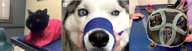
Methods of restraint used to provide a safe clinical examination in aggressive patients.
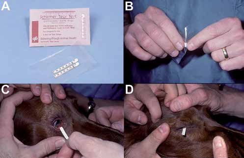
Evaluation of tear production: A) Schirmer tear-test strips for determining tear-film quantity (normal value in dogs is >15 mm/min; normal value in cats is between 8 and 15 mm/min); B) Bend the strip while it is still packaged to avoid contamination with skin oils, which can artificially alter test values; C) Place the strip in the lower conjunctival fornix; D) Close the eyelids for one minute, after which tear production can be read from the calibrated scale.
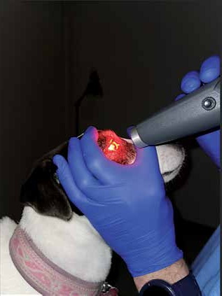
Testing the pupillary light reflex using chromatic light.
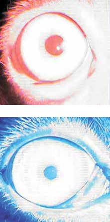
Normal pupillary response after testing with chromatic red and blue light.
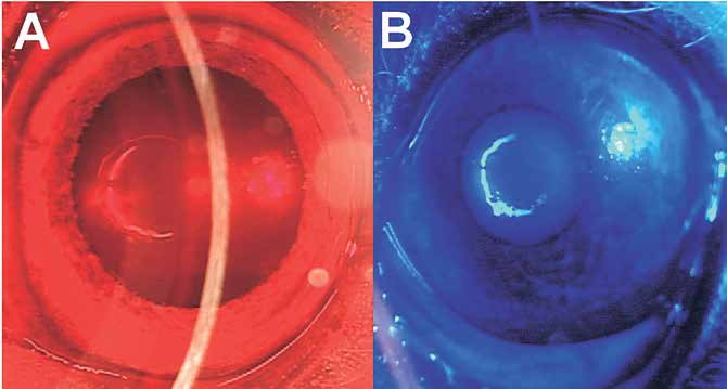
Retinal disease - chromatic-light testing (negative pupillary response to red light, positive response to blue light).
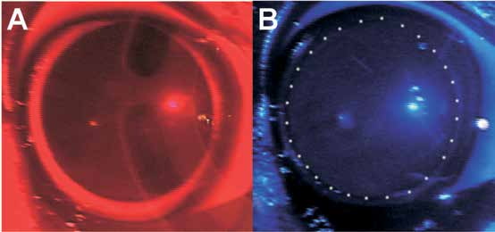
Optic nerve disease - chromatic-light testing (negative pupillary response to both red and blue light).
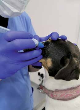
Tonometry - normal intraocular pressure in dogs and cats ranges from 15 to 25 mmHg.
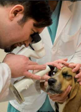
Slit-lamp examination of the eye.
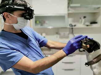
Examination using indirect ophthalmoscopy.
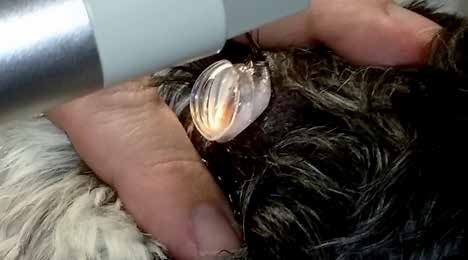
Gonioscopic examination of a dog to assess the patency of the anterior chamber drainage angles and predisposition to primary hereditary glaucoma.
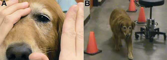
Evaluation of vision in a dog using the menace response and testing navigation around different objects (maze test).
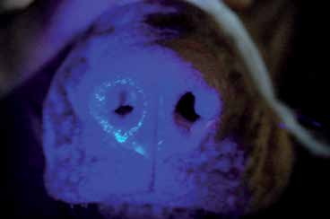
Jones test used to identify obstruction of the nasolacrimal ducts. Fluorescein is placed in both eyes, and within 5-10 minutes a trace of fluorescein should appear from both nostrils. In the dog shown, fluorescein appears from the right nostril, whereas the left remains unstained and dry; therefore, the nasolacrimal system of the left eye is not patent for some reason.
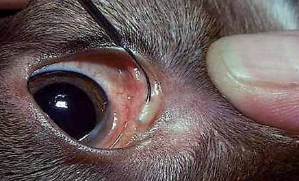
Flushing of the nasolacrimal duct to assess patency after application of topical anesthetic.
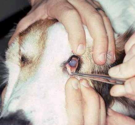
Evaluation of the inner surface of the third eyelid for the presence of a foreign body, parasitic organisms, and enlarged lymphoid follicles after application of topical anesthetic.
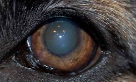
Dog with a corneal erosion that cannot be seen before fluorescein staining.
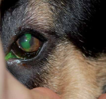
Corneal erosion that is readily visible after fluorescein staining.
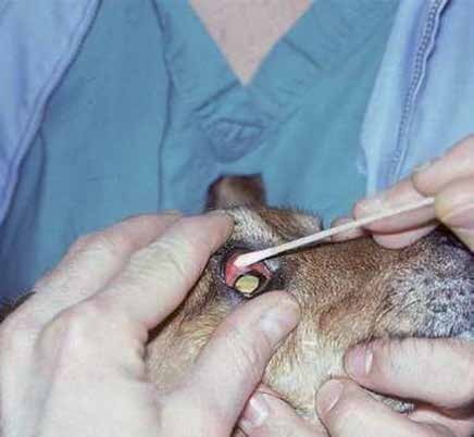
Sterile collection of a sample from the conjunctival surface for bacterial culture or PCR testing.
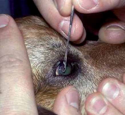
Collection of a sample for cytologic analysis using a Kimura spatula after application of topical anesthetic.
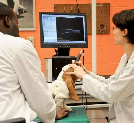
Ultrasound biomicroscopic examination of a dog - this technique enables detailed evaluation of the anterior chamber and aqueous humor drainage structures, allowing assessment of predisposition to primary hereditary glaucoma or detection of tumors of the anterior segment at a very early stage of development.
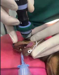
Ultrasound biomicroscopic examination of a dog. The dog is under general anesthesia because it has been prepared for surgery and the examination is being performed preoperatively. General anesthesia or sedation is usually not necessary for this examination.
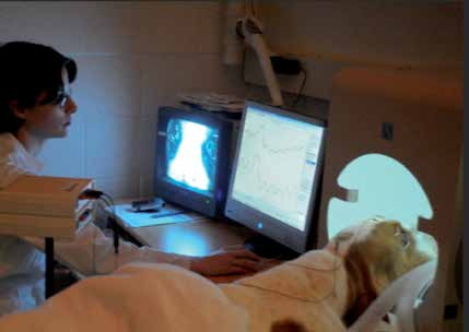
Electroretinography.
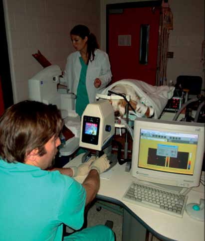
Contemporary techniques for examination of the optic nerve in a patient with hereditary glaucoma - GDx and OCT analysis of optic nerve structure.
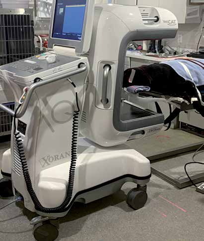
Contemporary techniques for examination of the orbit and brain using mobile computed tomography (CT) in a patient with an orbital tumor. This type of diagnostic imaging can also be used intraoperatively to confirm adequate resection of a mass.
Adapted from the Atlas of Ocular Diseases in Dogs and Cats by Dr. Siniša D. Grozdanić and colleagues. © Animal Eye Consultants of Iowa. The text and clinical images on this page are original content from the authors' own clinical practice and research, and may not be reproduced, distributed, or repurposed without permission.
Have questions about your pet's eyes?
Early evaluation gives us the most treatment options and the best chance of preserving vision.