
Chorioretinitis in Dogs
Canine chorioretinitis may be a primary ocular disorder or develop secondary to systemic disease. Causes listed in the source include neoplastic disease; fungal infections such as blastomycosis, histoplasmosis, cryptococcosis, and coccidioidomycosis; parasitic infections including Dirofilaria immitis, Onchocerca lupi, Toxocara canis, Leishmania infantum, Neospora caninum, and Toxoplasma gondii; viral diseases including herpesvirus and canine distemper; bacterial infections such as Brucella canis, Bartonella spp., and Leptospira spp.; vector-borne infection with Ehrlichia canis; autoimmune disorders including uveodermatologic syndrome and pancreatitis as listed in the source; and toxic and metabolic disease.
Fundic examination may show gray-white perivascular infiltrates from accumulation of inflammatory cells, gray hyporeflective areas in the tapetal region, hemorrhages of varying appearance and severity, retinal edema, and retinal detachment. Fungal infections often produce granular lesions. Inflammation can cause pigment proliferation in normally nonpigmented RPE beneath the tapetum, forming localized dark lesions. In chronic disease, retinal atrophy develops with tapetal hyperreflectivity and nontapetal depigmentation and pigment clumping.
Treatment
Diagnosis is established by ophthalmic examination. Complete blood count, serum biochemistry, and serologic testing for infectious disease help identify the cause. Treatment consists of topical and systemic anti-inflammatory medications and specific therapy for the underlying disease and complications.
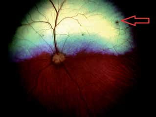
Chorioretinitis in a dog caused by blastomycosis. The red arrow indicates the lesion.
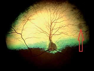
Chorioretinitis in a dog with toxoplasmosis. The red arrow indicates a retinal lesion.
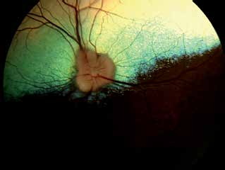
Chorioretinitis in a dog. Fundic lesions cannot be seen with indirect ophthalmoscopy.
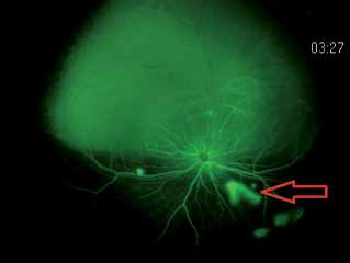
Chorioretinitis in the same dog as in the previous figure after fluorescein angiography. Chorioretinal exudative lesions become visible only with this diagnostic procedure, red arrow.
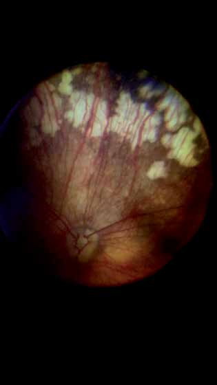
Autoimmune chorioretinopathy in which chronic inflammation causes progressive injury to the retinal pigment epithelium; depigmented zones represent damage.
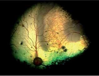
Chorioretinitis in a dog with chorioretinal scars, visible as dark hyperpigmented spots. The large gray area in the upper right fundus is a massive exudative lesion. The source identifies Cryptococcus spp. infection as the cause.
Related in-depth guide
Retinal Disorders
Our full guide, including treatment and surgical options
Adapted from the Atlas of Ocular Diseases in Dogs and Cats by Dr. Siniša D. Grozdanić and colleagues. © Animal Eye Consultants of Iowa. The text and clinical images on this page are original content from the authors' own clinical practice and research, and may not be reproduced, distributed, or repurposed without permission.
Think your pet may have chorioretinitis in dogs?
Early evaluation gives us the most treatment options and the best chance of preserving vision.