
Chorioretinal Lymphoma
Chorioretinal lymphoma is described as the second most common intraocular tumor in the source. Dogs with multicentric lymphoma most commonly show lymphadenopathy followed by ocular manifestations. The choroid and retina may also be involved, although less often than the anterior segment. Changes are usually bilateral. Posterior uveitis, retinal hemorrhage, altered tapetal coloration, papilledema, and retinal detachment may be seen.
Treatment
Diagnosis may be based on clinical and ophthalmic examination, cytologic analysis of aqueous humor and vitreous, and PCR. Complete blood count, serum biochemistry, lymph-node biopsy, thoracic radiography, and bone-marrow aspiration are recommended. Cats should be tested for FeLV, FIV, and FIP according to the source. Treatment consists of topical corticosteroids and systemic chemotherapy.
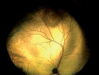
Chorioretinal lymphoma in a cat.
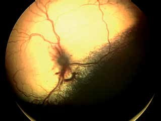
Chorioretinal lymphoma in a cat, with localized hemorrhages around the optic nerve head.
Related in-depth guide
Retinal Disorders
Our full guide, including treatment and surgical options
Adapted from the Atlas of Ocular Diseases in Dogs and Cats by Dr. Siniša D. Grozdanić and colleagues. © Animal Eye Consultants of Iowa. The text and clinical images on this page are original content from the authors' own clinical practice and research, and may not be reproduced, distributed, or repurposed without permission.
Think your pet may have chorioretinal lymphoma?
Early evaluation gives us the most treatment options and the best chance of preserving vision.