
Canine Atopic Dermatitis
Canine atopic dermatitis is a genetically predisposed, inflammatory, pruritic allergic skin disease with characteristic clinical signs, associated with production of IgE antibodies most commonly directed against environmental allergens. The primary clinical manifestation is pruritus, while primary skin lesions, when present, consist of erythema. Secondary lesions result from scratching, chronic skin inflammation, and concurrent secondary pyoderma and Malassezia dermatitis.
Skin findings include alopecia, excoriation, papules, pustules, dry broken hair, hyperpigmentation, scaling, and lichenification. These lesions generally occur in areas prone to scratching, including the face (muzzle and periocular region), concave surface of the pinna, dorsal and ventral surfaces of the paws, palmar aspect of the carpal and tarsal regions, axillae, abdomen, groin, and medial thighs. Initial signs most commonly develop by three years of age.
Canine atopic dermatitis may involve the eyelid skin and thereby cause qualitative or quantitative alterations in the tear film. Atopic dermatitis is also very frequently accompanied by atopic conjunctivitis.
Treatment
Diagnosis is based on clinical signs and exclusion of other pruritic dermatoses and is confirmed by allergy testing. Therapy is aimed at reducing pruritus and most commonly includes corticosteroids, cyclosporine, oclacitinib, monoclonal antibodies, and allergen-specific immunotherapy. Secondary bacterial and fungal infections of the skin and external ear canal must also be treated. Ocular therapy includes lacrimomimetics (tacrolimus or cyclosporine eye drops) and lubricants.
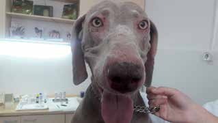
Atopic dermatitis. Alopecia and erythema around the eyes and nose.
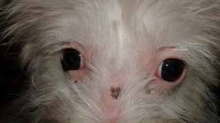
Atopic dermatitis. Periocular alopecia, edema, and erythema.
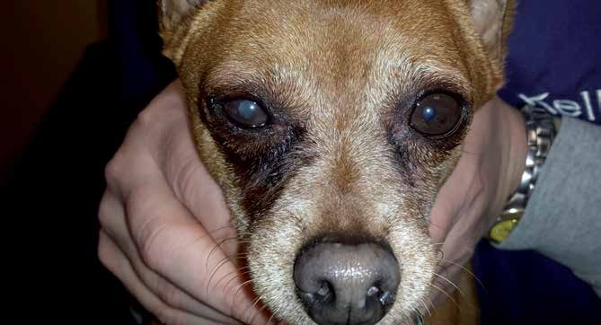
Atopic blepharitis and conjunctivitis in a dog resulting from ragweed pollen allergy. Changes are considerably more severe on the right side of the face.
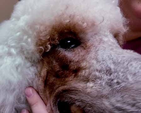
Atopic blepharitis and conjunctivitis with spastic entropion. Moist dermatitis developed as a result of increased tear secretion and wetting of the facial skin. Food allergy (chicken and beef) was identified in this patient.
Adapted from the Atlas of Ocular Diseases in Dogs and Cats by Dr. Siniša D. Grozdanić and colleagues. © Animal Eye Consultants of Iowa. The text and clinical images on this page are original content from the authors' own clinical practice and research, and may not be reproduced, distributed, or repurposed without permission.
Think your pet may have canine atopic dermatitis?
Early evaluation gives us the most treatment options and the best chance of preserving vision.