
Appearance of the Subalbinotic Fundus
Dogs and cats with blue irides or iris heterochromia and light-colored or merle/harlequin coats may have a subalbinotic fundus, a physiologic variation. The tapetal region is absent or poorly developed, and retinal pigment epithelium may lack pigment, allowing choroidal vessels to be seen. Choroidal vasculature against the white scleral background gives the fundus a characteristic tiger-striped appearance.
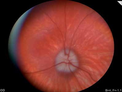
Subalbinotic canine fundus. Choroidal vasculature is clearly visible because of absence of the tapetum.
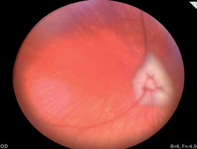
Subalbinotic canine fundus. Choroidal vasculature is clearly visible because of absence of the tapetum.
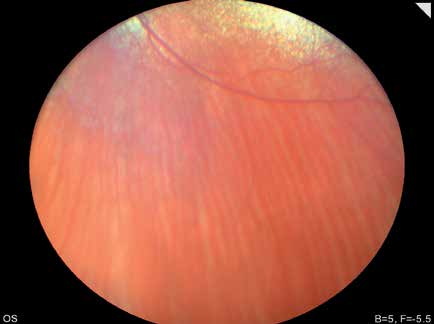
Subalbinotic feline fundus. A small yellow tapetal area is visible at the top of the image. Choroidal vessels are clearly visible in the nonpigmented nontapetal region.
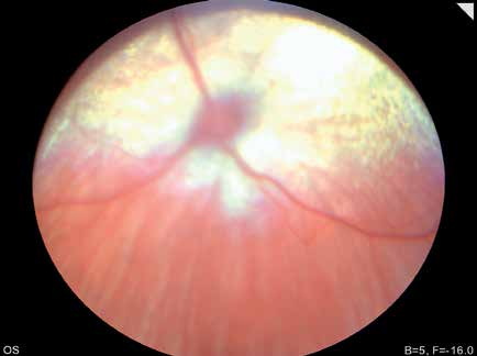
Subalbinotic feline fundus. A substantially larger yellow-white tapetal region is visible than in the previous figure. Choroidal vessels are clearly visible in the nonpigmented nontapetal region.
Related in-depth guide
Retinal Disorders
Our full guide, including treatment and surgical options
Adapted from the Atlas of Ocular Diseases in Dogs and Cats by Dr. Siniša D. Grozdanić and colleagues. © Animal Eye Consultants of Iowa. The text and clinical images on this page are original content from the authors' own clinical practice and research, and may not be reproduced, distributed, or repurposed without permission.
Think your pet may have appearance of the subalbinotic fundus?
Early evaluation gives us the most treatment options and the best chance of preserving vision.