
Appearance of the Feline Fundus
As in dogs, the feline fundus can be examined with direct or indirect ophthalmoscopy. The tapetal and nontapetal regions, optic nerve head, and retinal vessels can be identified. The feline tapetal region varies less in color and appearance than the canine tapetum. It is most often intensely yellow or yellow-green, triangular, and located dorsally. Cats with blue eyes may have partial or complete absence of the tapetum.
The nontapetal fundus is dark brown or black. In cats with light coat and eye color, it may be focally or diffusely nonpigmented, allowing visualization of choroidal vessels.
The optic nerve head appears as a small, round, nonmyelinated white or gray structure within the tapetal fundus. Unlike in dogs, retinal blood vessels do not cross the optic disc. Three pairs of arterioles and larger venules radiate from the optic nerve head.
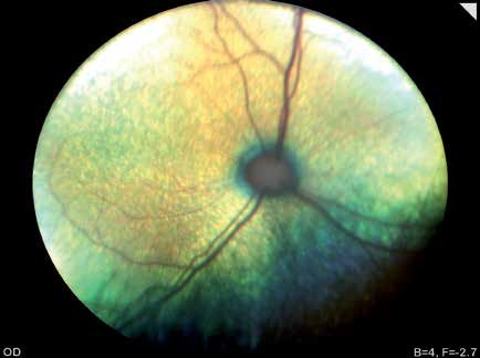
Normal feline fundus. There is a gradual transition from the yellow-green tapetal region to the pigmented nontapetal region, which appears dark blue to black. The optic nerve head is small, round, pink or gray, without the vascular ring seen in dogs. Three large veins radiate from the optic disc in a Mercedes-like configuration.
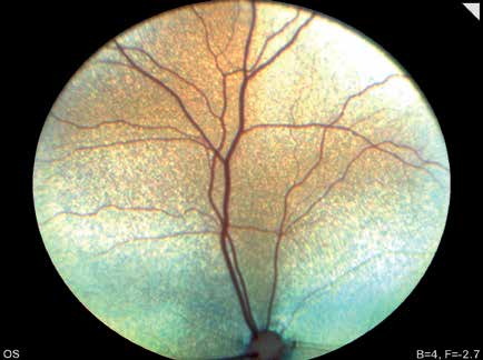
Normal variation of the feline fundus. The nontapetal region is not visible because only the tapetal portion was photographed. The optic nerve head is located near the bottom of the image and is small, round, and gray-pink.
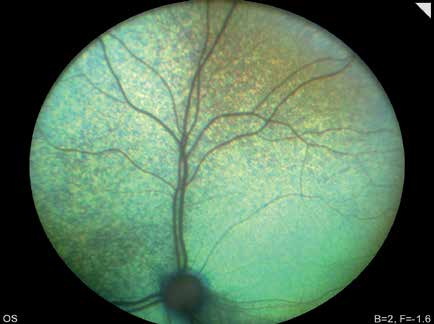
Normal variation of the feline fundus. Only the tapetal region is shown. The optic nerve head is small, round, gray, and located near the bottom of the image.
Related in-depth guide
Retinal Disorders
Our full guide, including treatment and surgical options
Adapted from the Atlas of Ocular Diseases in Dogs and Cats by Dr. Siniša D. Grozdanić and colleagues. © Animal Eye Consultants of Iowa. The text and clinical images on this page are original content from the authors' own clinical practice and research, and may not be reproduced, distributed, or repurposed without permission.
Think your pet may have appearance of the feline fundus?
Early evaluation gives us the most treatment options and the best chance of preserving vision.