
Appearance of the Canine Fundus
The canine fundus can be examined with direct or indirect ophthalmoscopy. Dogs show a very large range of normal fundic variation depending on age, breed, iris color, and coat color. During the first three to four months of life, the fundus of a puppy is bluish and later develops the appearance characteristic of the breed.
The canine fundus includes the tapetal region, nontapetal region, retinal blood vessels, and optic nerve head. The tapetal region is generally yellow or green but may also be blue, purple, or red-orange. Because the tapetum acts as a mirror that reflects light back through the retina, the underlying choroid and sclera cannot be seen in the tapetal region. The border between tapetal and nontapetal regions may be sharply defined or gradual and irregular. Dogs of the same breed generally have a similar tapetal appearance and color. Some dogs may lack a tapetum.
The nontapetal region is dark brown or black because pigment is present within the retinal pigment epithelium. In chocolate-colored dogs, the nontapetal region is often light brown or reddish brown. Retinal vessels are easily visible against the tapetal region but are more difficult to see in the nontapetal region.
The optic nerve, or optic disc/papilla, appears as a white or pink structure of variable shape and degree of myelination. It may be round, oval, triangular, or polygonal. An incomplete vascular ring surrounding a central depression is often visible. Three to four large veins and approximately 15-20 smaller arterioles extend from the optic nerve head.
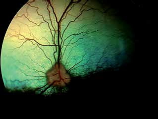
Canine fundus. Note shades of green, blue, and yellow in the tapetal region, the black nontapetal region, and the optic nerve head with prominent blood vessels and moderate myelination.
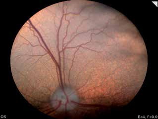
Normal variation of the canine fundus. Tapetum occupies most of the visible field and the nontapetal region is visible only at the lower edge as a darker pigmented area. A small, dark-gray, round physiologic depression is present in the center of the optic nerve head.
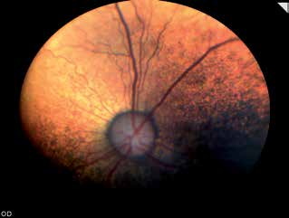
Normal variation of the canine fundus. The nontapetal region is larger than in the previous figure. The tapetal region has orange coloration and the optic nerve head is round.
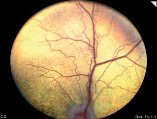
Normal variation of the canine fundus. Only a light tapetal region with prominent vascularization is visible. Larger veins and thinner arteries can be clearly distinguished.
Related in-depth guide
Retinal Disorders
Our full guide, including treatment and surgical options
Adapted from the Atlas of Ocular Diseases in Dogs and Cats by Dr. Siniša D. Grozdanić and colleagues. © Animal Eye Consultants of Iowa. The text and clinical images on this page are original content from the authors' own clinical practice and research, and may not be reproduced, distributed, or repurposed without permission.
Think your pet may have appearance of the canine fundus?
Early evaluation gives us the most treatment options and the best chance of preserving vision.