
Anterior Uveitis in Cats
Uveitis is inflammation of the uveal tract, which consists of the iris, ciliary body, and choroid. It is common in cats because these tissues are highly vascularized and immunologically active. Anterior uveitis includes iritis, cyclitis, and iridocyclitis, whereas posterior uveitis includes choroiditis and chorioretinitis.
Uveitis can occur independently or secondary to disease of the lens, cornea, or sclera. It may be a primary ocular disorder or may occur as a consequence of systemic neoplastic, infectious, autoimmune, toxic, or metabolic disease. Infectious causes listed in the source include FeLV, FIP, Toxoplasma gondii, Cryptococcus neoformans, and Bartonella spp. The source states that more than 50% of feline uveitis cases are associated with systemic disease. Because uveitis can cause loss of vision or may be a clinical sign of a life-threatening systemic illness, determining the cause is very important.
Feline anterior uveitis may affect one or both eyes and is markedly painful, producing blepharospasm and photophobia. Epiphora, ocular discharge, and third eyelid protrusion may be present. Ocular redness, conjunctival hyperemia, scleral injection, and corneal neovascularization may occur. The eye can appear cloudy because of corneal edema, keratic precipitates consisting of inflammatory cells deposited on the corneal endothelium, and fibrin within the anterior chamber.
Hypopyon or hyphema may develop. The iris becomes darker, and iris neovascularization (rubeosis iridis) may occur. The pupil is constricted, with possible anisocoria and dyscoria. Complications include synechiae, secondary glaucoma, cataract, and iris bombe with anterior ballooning of the iris toward the corneal endothelium.
Treatment
Diagnosis is established by ophthalmic examination. In bilateral uveitis, systemic causes should be excluded. Recommended investigations include a complete blood count and serum biochemistry profile, thoracic radiography, abdominal ultrasonography, cytologic examination of lymph nodes particularly when enlarged, and serologic testing for infectious disease. Treatment consists of topical and systemic anti-inflammatory medications together with specific treatment directed at the identified cause and any complications.
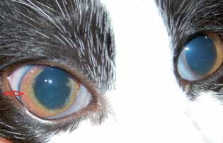
Bilateral uveitis in a cat diagnosed with FIP. Keratic precipitates and iris neovascularization, indicated by the red arrow, are visible.
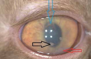
Uveitis in a cat diagnosed with FIP. A cloudy, edematous cornea, hypopyon indicated by the red arrow, fibrin deposits on the corneal endothelium indicated by the black arrow, and iris neovascularization are visible. The blue arrow indicates synechiae between the iris and lens.
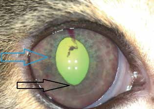
Uveitis in a cat. Uveal ectropion is present; the black arrow indicates the everted iris surface. Numerous iris nodules represent accumulations of inflammatory cells, indicated by the blue arrow. Blood within the anterior chamber and iris neovascularization are visible.
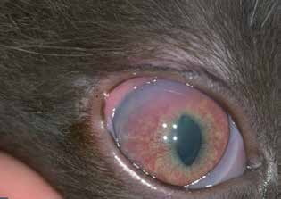
Uveitis in a cat. Miosis and dyscoria, corneal edema and vascularization, and iris neovascularization are visible.
Adapted from the Atlas of Ocular Diseases in Dogs and Cats by Dr. Siniša D. Grozdanić and colleagues. © Animal Eye Consultants of Iowa. The text and clinical images on this page are original content from the authors' own clinical practice and research, and may not be reproduced, distributed, or repurposed without permission.
Think your pet may have anterior uveitis in cats?
Early evaluation gives us the most treatment options and the best chance of preserving vision.