
Anterior Lens Luxation
In dogs and cats, the lens can dislocate from its normal position in the patellar fossa either anteriorly into the anterior chamber or posteriorly into the vitreous. Luxation may be inherited, as in primary bilateral lens luxation associated in the source with an ADAMTS gene mutation in terriers, especially Jack Russell Terriers, as well as Border Collies, Chow Chows, and mixed breeds derived from these breeds. Clinical disease most commonly develops between three and six years of age. The genetic defect weakens the zonular ligaments supporting the lens, predisposing the lens to dislocation. Primary lens luxation is bilateral but not symmetric.
Lens luxation can also accompany hereditary ocular abnormalities in many breeds or develop secondarily to abnormal ocular development, degenerative processes, trauma, uveitis, cataract, or glaucoma.
With anterior displacement, the lens may push the iris anteriorly while remaining in the posterior chamber or may move completely into the anterior chamber. In either case, anterior chamber depth decreases. Clinical consequences include marked discomfort, corneal edema due to contact between the lens capsule and corneal endothelium, glaucoma due to impaired aqueous flow, and uveitis caused by abnormal contact between the lens, vitreous, and anterior uveal structures. If cataract did not cause the original lens dislocation, luxation itself can result in cataract, making the primary process difficult to determine. Careful retinal examination is important because the source states that retinal damage and early retinal detachment occur in nearly 50% of clinical cases.
Treatment
Ophthalmic examination may reveal the lens within the anterior chamber, a crescent-shaped aphakic reflection behind the lens margin, corneal edema, elongated ciliary processes, vitreous in the anterior chamber, cataract, and glaucoma. Ultrasonography can confirm the diagnosis when the cornea is opaque. Medical management may attempt to move the lens from an anterior to a posterior position and maintain miosis to keep it trapped in the posterior segment. Surgical intracapsular lens extraction or phacoemulsification is described as the most successful treatment.
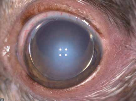
Primary anterior lens luxation in a terrier. Corneal edema is visible.
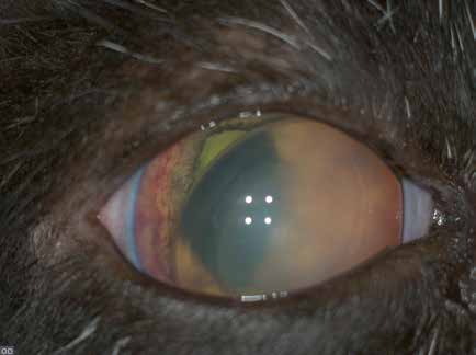
Anterior lens luxation in a cat secondary to uveitis.
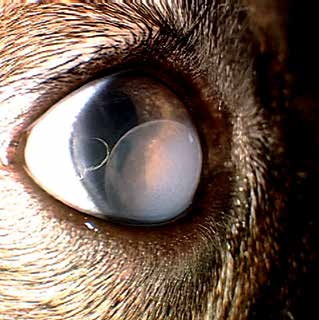
Anterior lens luxation in a dog.
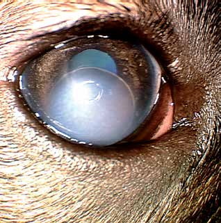
Anterior lens luxation in the same dog as in the previous figure, viewed from another angle.
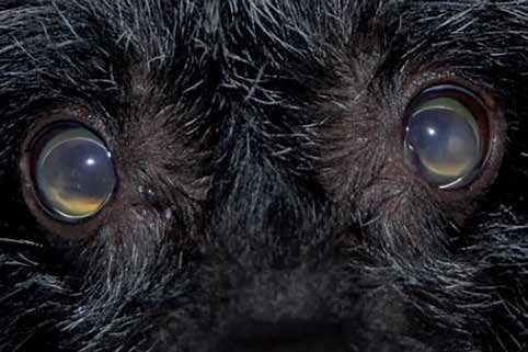
Bilateral anterior lens luxation in a dog.
Adapted from the Atlas of Ocular Diseases in Dogs and Cats by Dr. Siniša D. Grozdanić and colleagues. © Animal Eye Consultants of Iowa. The text and clinical images on this page are original content from the authors' own clinical practice and research, and may not be reproduced, distributed, or repurposed without permission.
Think your pet may have anterior lens luxation?
Early evaluation gives us the most treatment options and the best chance of preserving vision.