
Anterior Chamber Lipidosis
Anterior chamber lipidosis is the presence of varying amounts of lipoproteins in the anterior chamber. Vascular endothelium of iris vessels and nonpigmented ciliary epithelium form a blood-ocular barrier that normally prevents larger blood particles from entering the anterior chamber. This barrier fails during uveitis, allowing lipids to enter, particularly in dogs with hyperlipidemia.
In dogs with hyperlipidemia, anterior chamber lipidosis may sometimes develop primarily without preceding uveitis, and may then itself lead to secondary uveitis. Corneal lipid accumulation and/or lipemia of the retinal vasculature may accompany the condition. In dogs and cats, it appears as whitish ocular cloudiness that may resemble corneal edema or cataract. Miniature Schnauzers are predisposed. The condition has also been diagnosed in cats.
Treatment
Diagnosis is established by ophthalmic examination. Complete blood count and serum biochemistry are performed, with measurement of cholesterol and triglycerides. Further testing investigates the underlying metabolic disorder, including liver, kidney, pancreatic, and thyroid function. If uveitis caused the lipidosis, the cause of the uveitis should be determined. Treatment includes a low-fat diet and management of the underlying cause of hyperlipidemia or uveitis. Steroidal and nonsteroidal anti-inflammatory eye drops are used to control uveitis.
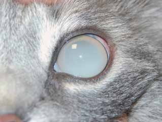
Anterior chamber lipidosis in a cat.
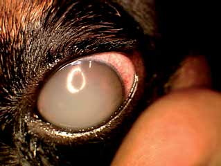
Anterior chamber lipidosis in a dog.
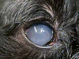
Anterior chamber lipidosis in a dog. Intraocular structures are not visible because of the lipid.
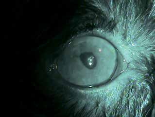
Infrared image of the eye of a dog. Infrared imaging makes internal ocular structures visible despite anterior chamber lipidosis.
Adapted from the Atlas of Ocular Diseases in Dogs and Cats by Dr. Siniša D. Grozdanić and colleagues. © Animal Eye Consultants of Iowa. The text and clinical images on this page are original content from the authors' own clinical practice and research, and may not be reproduced, distributed, or repurposed without permission.
Think your pet may have anterior chamber lipidosis?
Early evaluation gives us the most treatment options and the best chance of preserving vision.