
Vitreoretinal Surgery for Dogs and Cats
Advanced treatment for retinal detachment, giant retinal tears, and vitreoretinal disease at the Animal Eye Consultants of Iowa
What is a retinal detachment?
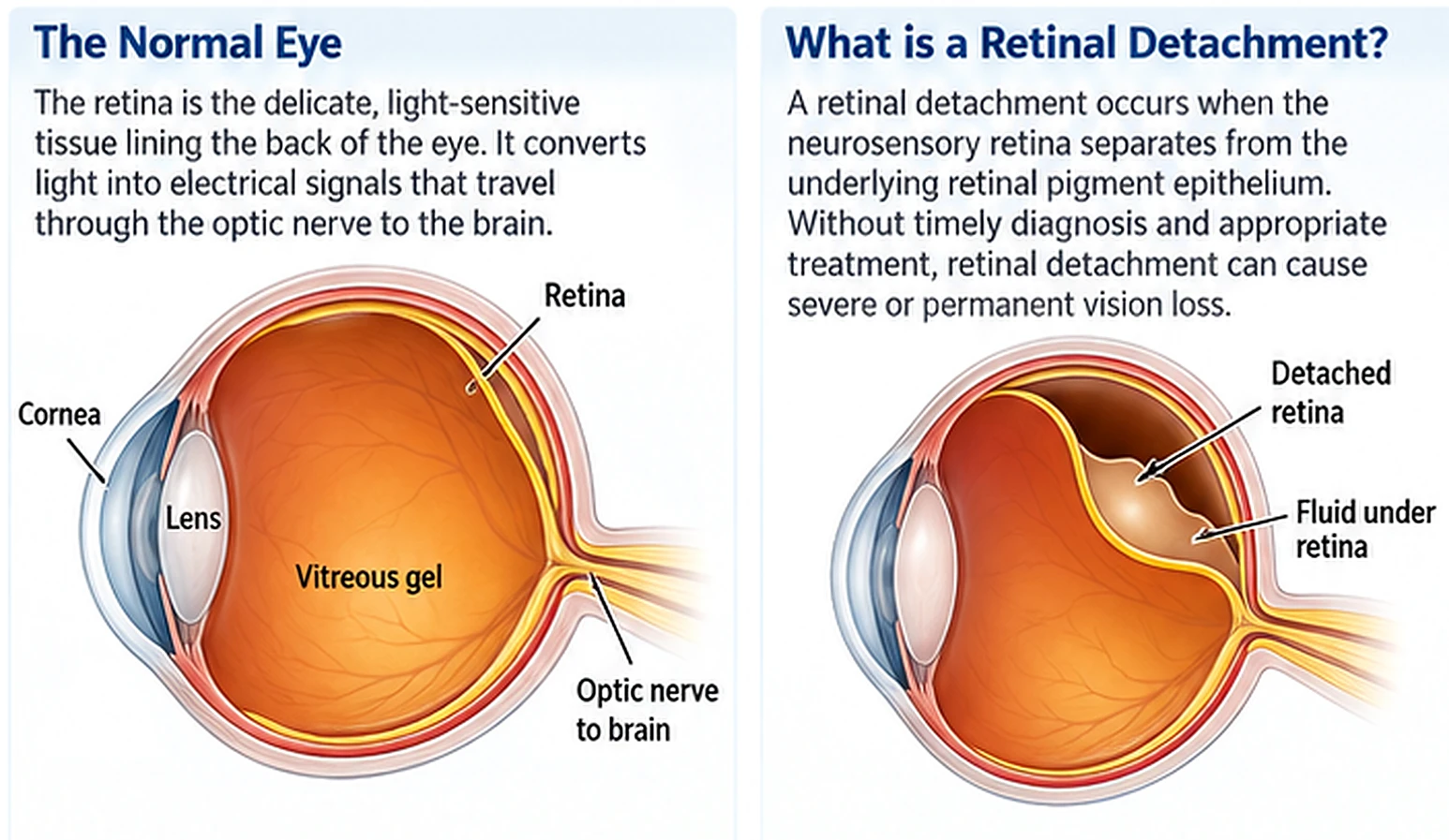
The retina is the delicate, light-sensitive tissue lining the back of the eye. It converts light into electrical signals that travel through the optic nerve to the brain. A retinal detachment occurs when the neurosensory retina separates from the underlying retinal pigment epithelium. Without timely diagnosis and appropriate treatment, retinal detachment can cause severe or permanent vision loss.
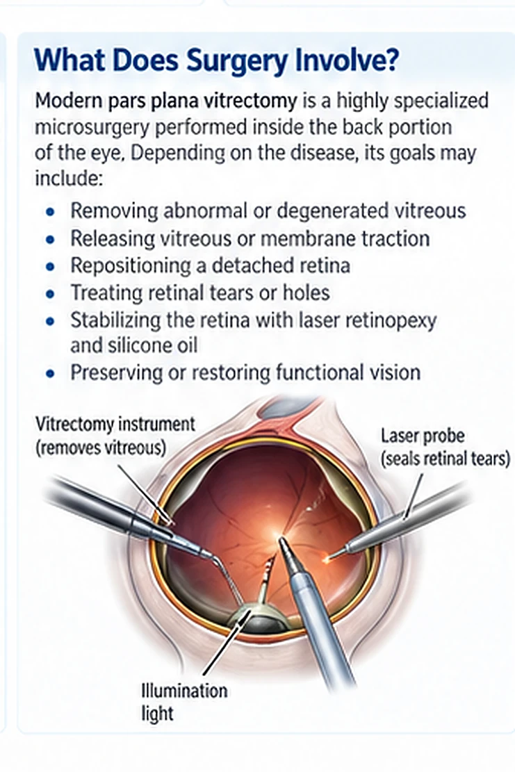
Vitreoretinal surgery is highly specialized microsurgery performed inside the back portion of the eye. Depending on the disease, its goals may include:
- Removing abnormal or degenerated vitreous
- Releasing vitreous or membrane traction on the retina
- Repositioning a detached retina
- Treating retinal tears or holes
- Stabilizing the retina with laser retinopexy and silicone oil
- Preserving or restoring functional vision
Modern pars plana vitrectomy has made surgical repair possible in selected dogs and cats that previously would have remained permanently blind.
Not every retinal detachment requires surgery
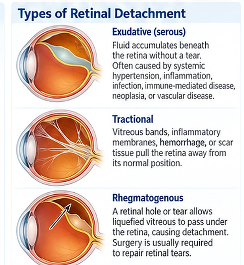
Retinal detachments are classified according to their underlying mechanism.
Exudative or serous retinal detachment
In an exudative detachment, fluid accumulates beneath the retina without a retinal tear. Causes can include systemic hypertension, intraocular inflammation, infectious disease, immune-mediated disease, neoplasia, and choroidal or vascular disease. These detachments are generally treated by identifying and controlling the underlying disease. In cats, systemic hypertension is a particularly important cause of bullous retinal detachment. When treatment begins before irreversible retinal injury occurs, the retina may return toward its normal position and vision may improve.
Tractional retinal detachment
Tractional detachment occurs when vitreous bands, inflammatory membranes, hemorrhage, or scar tissue pull the retina away from its normal position. Some tractional detachments require surgery, particularly when traction is progressive or accompanied by retinal tearing.
Rhegmatogenous retinal detachment
A rhegmatogenous detachment develops when liquefied vitreous passes through a retinal hole or tear. In dogs, these tears are frequently extensive and may become giant retinal tears or retinal dialyses. Medical treatment cannot seal a retinal tear or eliminate mechanical vitreous traction; surgical reattachment is therefore usually required when restoration of vision is the goal. Some eyes, particularly after cataract surgery or severe intraocular inflammation, may develop both tractional and rhegmatogenous components of retinal detachment.
Conditions that may be treated surgically
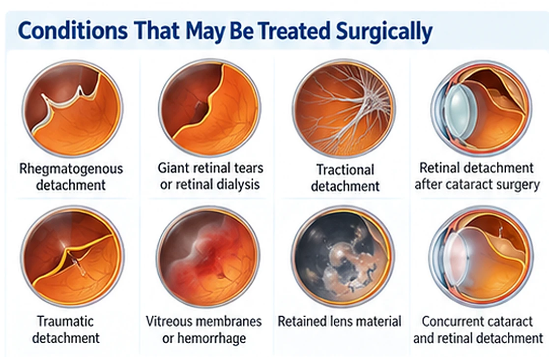
Vitreoretinal surgery may be considered for selected dogs and cats with:
- Rhegmatogenous retinal detachment
- Giant retinal tears or retinal dialysis
- Tractional or combined tractional–rhegmatogenous detachment
- Retinal detachment associated with vitreous degeneration
- Retinal detachment following cataract or lens surgery
- Retinal tears associated with lens-induced inflammation
- Traumatic retinal tears or detachments
- Vitreous membranes that distort or pull on the retina
- Selected cases of retained lens material, vitreous hemorrhage, or intraocular scarring
- Concurrent cataract and surgically repairable retinal detachment
Canine risk factors for retinal detachment described in the literature include inherited vitreous abnormalities, retinal dysplasia, cataract and lens-induced uveitis, previous cataract surgery, lens luxation, trauma, retinal cysts, and Collie eye anomaly.
Signs of retinal detachment
A pet with retinal detachment may:
- Suddenly bump into furniture or walls
- Become hesitant on stairs
- Have difficulty finding food, toys, or doorways
- Appear disoriented in unfamiliar surroundings
- Lose vision in dim light before bright-light vision
- Develop one or two persistently dilated pupils
- Show a sudden change in eye color or fundus reflection
- Develop redness, cloudiness, hemorrhage, or discomfort when inflammation or glaucoma is also present
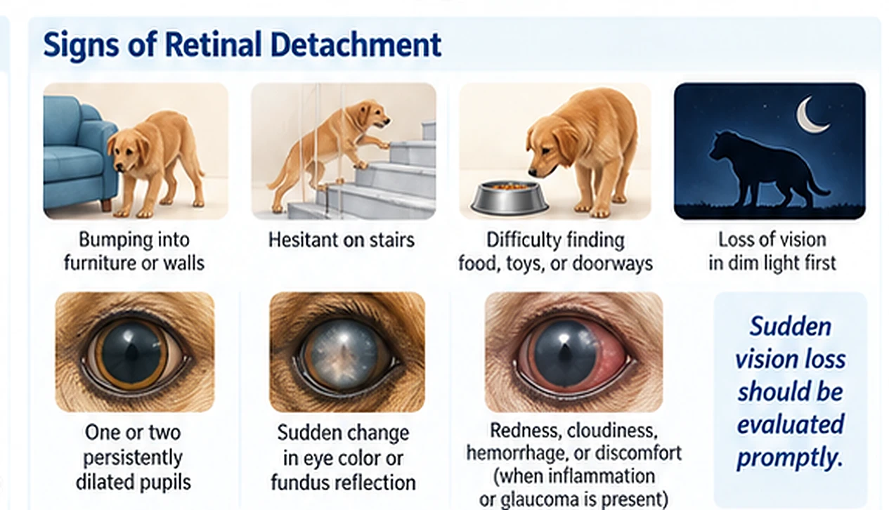
Sudden vision loss should be evaluated promptly. The examination must determine not only whether the retina is detached, but also whether a retinal tear is present and whether the detachment is surgical or potentially responsive to medical therapy.
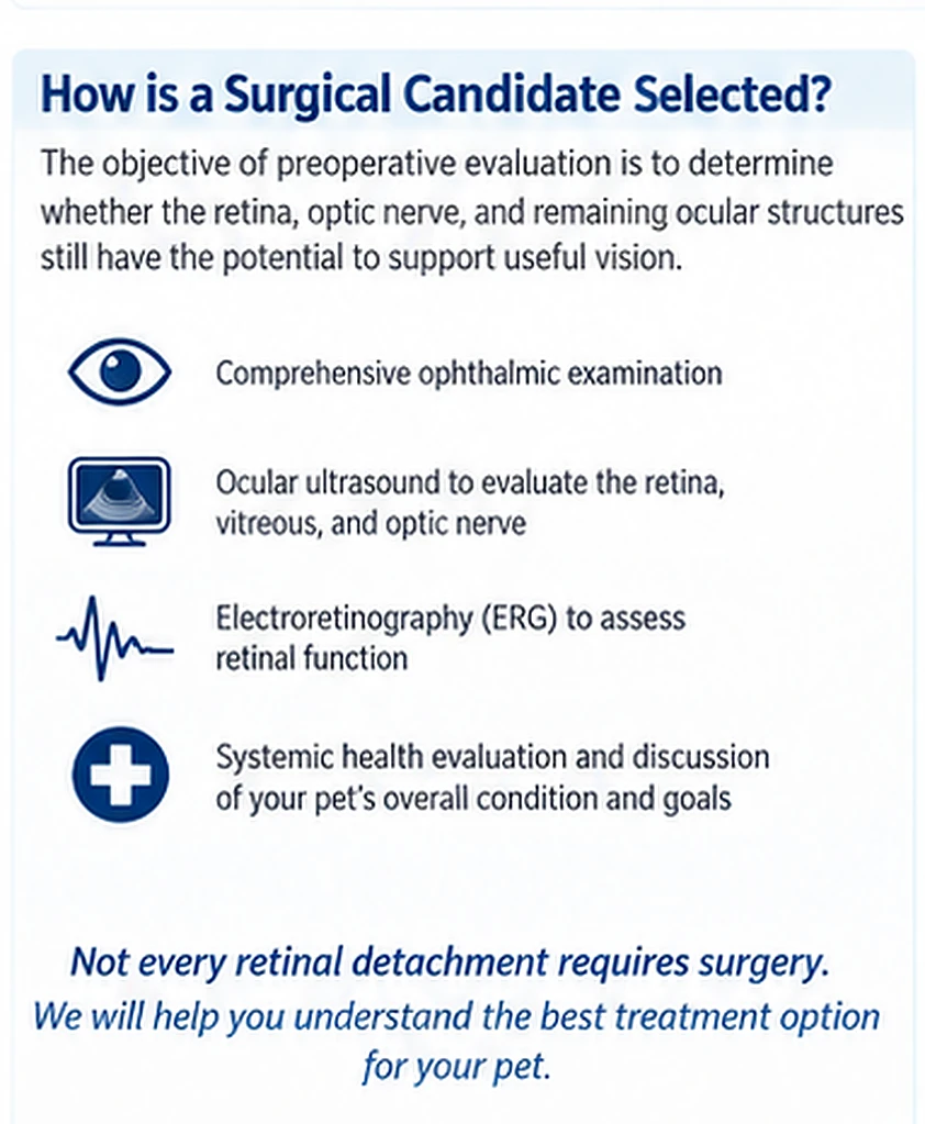
How is a surgical candidate selected?
The objective of preoperative evaluation is not simply to determine whether the retina can be physically reattached. The more important question is whether the retina, optic nerve, and remaining ocular structures retain sufficient function to support useful postoperative vision. Evaluation may include:
- Complete ophthalmic examination
- Menace-response assessment
- Dazzle-reflex assessment
- Direct and consensual pupil-light reflexes
- Chromatic pupil-light-reflex testing in selected patients
- Detailed assessment of retinal architecture and blood vessels
- Indirect ophthalmoscopy
- Ocular ultrasonography
- Electroretinography
- Intraocular-pressure measurement
- Examination for cataract, lens instability, hemorrhage, inflammation, and previous surgical changes
- Blood-pressure measurement and systemic testing when an underlying disease is suspected
Findings associated with a more favorable prognosis
The strongest clinically useful favorable findings include:
- A detectable menace response
- A detectable dazzle reflex
- A recordable electroretinogram
- Relatively healthy retinal tissue architecture
- Preserved retinal vasculature and perfusion
- A partial rather than extensively contracted detachment
- A surgically manageable tear or area of traction
- Controlled intraocular pressure
- Minimal intraocular inflammation
- Absence of advanced optic-nerve damage
In a study of 275 operated canine eyes, postoperative vision was documented in 87.7% of eyes with a preoperative menace response versus 69.3% without one, and in 78.5% of eyes with a dazzle reflex versus 58.9% without one. Vision returned in 90.4% of eyes graded as having good retinal architecture, 69.3% with fair architecture, and 50.8% with poor architecture — findings that show poor retinal appearance reduces the probability of vision recovery but does not necessarily make recovery impossible.
The role of electroretinography
Electroretinography objectively measures retinal electrical activity and is one of the most useful preoperative prognostic tests. In a study of 40 canine eyes, all 12 eyes with normal or attenuated recordable ERGs regained normal vision, and normal vision returned in 18 of 28 eyes — 64% — with flat preoperative ERGs. A recordable ERG is therefore a favorable prognostic finding, while a flat ERG lowers the expected probability of visual recovery but does not prove that recovery is impossible.
Duration of retinal detachment
Earlier recommendations frequently used one month as a practical cutoff for a favorable prognosis. More recent evidence indicates that duration should not be used as the sole reason to accept or reject a patient. In one series, the proportion of eyes regaining or maintaining vision declined from 77% for detachments estimated at 0–14 days to 66% for detachments estimated at 28–60 days, though duration was not statistically associated with visual outcome in the overall analysis. Earlier surgery is generally preferable because prolonged separation can produce progressive photoreceptor, retinal pigment epithelial, vascular, and neural changes — but an eye should be evaluated individually rather than excluded solely because the exact duration is uncertain.
Age and pupillary light reflexes
Age alone should not be used to determine candidacy — in the largest canine study, age was not significantly associated with postoperative vision. A conventional positive pupillary light reflex is also not an independent guarantee of visual recovery: vision returned or was maintained in 74.5% of eyes with a positive PLR and 73.1% of eyes with a negative PLR. The menace response, dazzle reflex, retinal architecture, ERG, and optic-nerve status are more informative when considered together.
Guarded and poor surgical candidates
Cases requiring a more guarded prognosis
Surgery may still be possible, but the prognosis is more guarded when there is:
- Longer or uncertain duration of detachment
- Poor or difficult-to-assess retinal architecture
- Absent dazzle responses
- A flat electroretinogram
- Previous intraocular surgery
- Cataract, aphakia, or lens instability
- Retained lens material
- Mild or moderate intraocular inflammation
- Extensive vitreous membranes or proliferative changes
- Multiple retinal tears
- Partial retinal nonperfusion
- Microphthalmos or other developmental abnormalities
- Previous retinal reattachment surgery
No single guarded finding necessarily excludes surgery. The cumulative condition of the retina, optic nerve, cornea, lens, and intraocular-pressure control determines the recommendation.
Findings commonly associated with a poor prognosis
Surgery may offer little probability of visual restoration when there is:
- Uncontrolled glaucoma or severe buphthalmos
- Advanced optic-nerve atrophy or glaucomatous cupping
- Severe diffuse retinal degeneration or nonperfusion
- Severe retinoschisis
- Endophthalmitis or uncontrolled intraocular infection
- Severe active intraocular inflammation
- Extensive retinal fibrosis or a tightly contracted, cylindrical retina
- Multiple large radial tears that cannot be stabilized
- Complete choroidal detachment
- Scleral rupture or necrosis
- Extensive proliferative vitreoretinopathy
- Evidence that blindness is caused primarily by optic-nerve or central neurologic disease
What happens during vitreoretinal surgery?
Vitreoretinal surgery is performed under general anesthesia using an operating microscope, specialized posterior-segment viewing equipment, and microsurgical instruments introduced through small openings in the sclera.
Pars plana vitrectomy
The surgeon removes liquefied or abnormal vitreous and releases tractional bands or membranes from the retinal surface. Peripheral vitreous that continues to pull on the tear is carefully removed.
Retinal unfolding and repositioning
A temporary heavy perfluorocarbon liquid may be placed over the retina. Its weight helps unfold and reposition the detached retina while displacing subretinal fluid.
Treatment of tears
Laser retinopexy is applied around retinal tears or structurally weak areas. Laser treatment creates a chorioretinal adhesion that helps prevent fluid from passing through the retinal break.
Silicone-oil tamponade
Silicone oil replaces the vitreous and supports the retina after surgery. It is particularly useful in veterinary patients because dogs and cats cannot reliably maintain the prolonged postoperative head positioning required for gas tamponade. Silicone oil is often retained for an extended period in veterinary patients — removal may be considered in selected eyes, but it can be associated with recurrent detachment or retinal slippage.
Small-gauge vitrectomy
Current instrumentation permits smaller surgical ports than the original 20-gauge techniques. A 2025 study comparing 25- and 27-gauge canine pars plana vitrectomy reported 100% immediate anatomical success with both systems, with visual success of 85.0% for 25-gauge and 78.9% for 27-gauge procedures, and no significant difference in overall surgical time, visual outcome, or postoperative complication rates. The 27-gauge procedure had faster trocar placement but more intraoperative incidents during the vitrectomy and reattachment phase.
Cataract and retinal-detachment surgery
Cataracts may coexist with retinal detachment or develop after successful retinal surgery.
Combined cataract and retinal surgery
When a cataract prevents adequate visualization or access to the retina, phacoemulsification and retinal reattachment surgery are performed during the same anesthetic procedure. In a study limited to Siberian Huskies with cataract and preoperative retinal detachment, all 17 eyes were anatomically reattached immediately after surgery, and functional vision was maintained or restored in 15 of 17 eyes — 88.2%. Silicone-oil migration into the anterior chamber occurred in 47.1%, and corneal endothelial decompensation and glaucoma each occurred in 17.6%. These results are encouraging but represent a small, breed-specific retrospective population and should not be interpreted as a guaranteed success rate for every patient.
Cataract surgery after retinal reattachment
Cataract formation or progression is a recognized complication of retinal reattachment surgery. In one study of dogs undergoing cataract surgery after previous retinal repair, immediate visual improvement occurred in 39 of 41 eyes — 95.1%. Among eyes available for follow-up, vision was maintained in 93.9% at one month, 85.2% at three months, 77.3% at six months, and 60% at 12 months. Glaucoma, corneal ulceration, fibrin formation, and anterior migration of silicone oil were the most frequent complications. Cataract surgery can therefore be considered when a successfully reattached retina remains functional but a cataract subsequently blocks vision.
Expected results in dogs
Anatomical success and visual success are different outcomes. A retina may be successfully repositioned yet fail to restore useful vision if irreversible photoreceptor, retinal, or optic-nerve injury occurred before surgery. Published canine studies have reported immediate anatomical reattachment in approximately 90% to 100% of operated eyes, and postoperative visual success in approximately 72% to 92% of operated eyes. The variation reflects differences in patient selection, retinal pathology, duration, surgical technique, definitions of vision, postoperative complications, and follow-up.
In one study of 168 eyes, 155 — 92% — were visual after surgery, and most of these eyes had been nonvisual before surgery. In a larger series of 275 eyes, immediate anatomical success was 98%, vision was maintained or restored in 72% of eyes, and vision returned an average of 18.5 days after surgery. Among eyes that regained vision, 71.7% remained visual at the last examination, with an average follow-up of approximately 550 days. These studies demonstrate that useful vision can return in a substantial proportion of carefully selected blind dogs, but no individual patient can be guaranteed to regain or permanently retain vision.
Vitreoretinal surgery in cats
Most feline retinal detachments are exudative and are frequently associated with systemic hypertension. These cases are usually managed medically by controlling blood pressure and the underlying disease.
Cats can also develop giant retinal tears, particularly with trauma or vitreous degeneration. A study from Animal Eye Consultants of Iowa reviewed 99 cats with retinal detachment: sixteen cats — 16.1% — had a giant retinal tear, and trauma, vitreous degeneration, and male sex were associated with giant-tear development. Three cats underwent vitreoretinal reattachment with silicone-oil tamponade, and all three surgically treated cats regained vision with a median follow-up of 565 days; glaucoma or ocular hypertension developed in two of the three operated cats. These results demonstrate that feline vitreoretinal surgery is technically possible and can restore vision.
Feline surgery presents additional technical challenges because the feline lens is proportionally large, limiting access to the posterior segment — feline reattachment surgery is therefore always combined with lens removal surgery.
Possible complications
Vitreoretinal surgery is complex and requires long-term monitoring. Potential complications include:
- Silicone-oil migration into the anterior chamber
- Increased intraocular pressure or glaucoma
- Cataract formation or progression
- Corneal ulceration
- Corneal endothelial degeneration or edema
- Uveitis or fibrin formation
- Retinal degeneration
- Recurrent retinal detachment
- Proliferative vitreoretinopathy
- Intraocular hemorrhage
- Infection
- Lens instability
- Need for additional surgery
- Permanent vision reduction or loss
In one 275-eye series, the most frequently documented complications were anterior silicone-oil migration, corneal ulceration, glaucoma, and cataract formation.
Does silicone oil change vision?
Silicone oil changes the refractive properties of the eye. A study of 63 silicone-oil– filled canine eyes found hyperopic refractive error in phakic, pseudophakic, and aphakic eyes; in aphakic eyes, the oil reduced the severe hyperopia normally associated with absence of the lens, producing a relative shift toward myopia compared with untreated aphakia. This means a successfully operated pet may have useful functional vision without having the same optical focus as a normal eye. Most dogs adapt well to moderate refractive differences.
Recovery and follow-up
After surgery, patients require close monitoring of:
- Retinal attachment
- Intraocular pressure
- Inflammation
- Corneal health
- Lens clarity
- Silicone-oil position
- Visual responses
Postoperative care generally includes topical medications, systemic medications when indicated, an Elizabethan collar, restricted activity, and scheduled examinations.
Vision may return immediately, but more commonly improves gradually over several days or weeks — in one large study, the average time to documented vision recovery was approximately 18.5 days. Some pets initially show only bright-light navigation or object avoidance before developing more consistent functional vision, and in some cases it can take up to four months before functional vision returns.
Owners should seek prompt examination if they observe redness, squinting, discharge, corneal cloudiness, enlargement of the eye, apparent pain, or deterioration in vision.
Frequently asked questions
Can a completely blind dog regain vision?
Yes. Many dogs included in published surgical studies were blind before surgery, and a substantial proportion regained functional vision. The probability depends on retinal architecture, ERG activity, visual reflexes, optic-nerve health, intraocular pressure, and the underlying cause of detachment.
Does a flat ERG mean that surgery cannot work?
No. A recordable ERG is favorable, but 64% of eyes with a flat preoperative ERG regained normal vision in one prospective study. A flat ERG should be interpreted as a guarded — not automatically hopeless — finding.
Is an older pet automatically a poor candidate?
No. Age was not independently associated with postoperative vision in the largest canine study. General health and the condition of the retina, optic nerve, cornea, and remainder of the eye are more important.
Does the surgery have to be performed within one month?
Earlier evaluation and treatment are preferred, but one month is not an absolute evidence-based cutoff. Duration should be considered together with retinal appearance, ERG findings, visual reflexes, intraocular pressure, and optic-nerve health.
Can cats undergo retinal reattachment surgery?
Yes, in selected cases involving a retinal tear or surgically correctable traction, cats can undergo retinal reattachment surgery.
Can cataract surgery be performed at the same time?
Yes, when cataract prevents retinal visualization or surgical access. Combined phacoemulsification and pars plana vitrectomy may avoid two separate anesthetic events, but the recommendation depends on lens stability, retinal configuration, inflammation, and glaucoma risk. In cats, retinal reattachment surgery is always performed in combination with lens removal.
Is every retinal detachment an emergency?
Yes, retinal detachments are emergency conditions. Sudden vision loss warrants urgent evaluation. Exudative detachments may require immediate treatment of systemic hypertension or inflammation, whereas retinal tears require prompt assessment for possible surgery — the treatment depends on the mechanism of detachment.
Scientific references
Incidence of and treatment strategies for giant retinal tears in cats with retinal detachment
Grozdanić SD, Luzhetskii S, Lazić T. Canadian Veterinary Journal. 2026;67(6):670-678.
Management of giant retinal tears in dogs
Vainisi SJ, Packo KH. Journal of the American Veterinary Medical Association. 1995;206:491-495. doi:10.2460/javma.1995.206.04.491.
Canine retinal surgery
Vainisi SJ, Wolfer JC. Veterinary Ophthalmology. 2004;7:291-306. doi:10.1111/j.1463-5224.2004.04049.x.
Outcome of retinal reattachment surgery in dogs: a retrospective study of 145 cases
Steele KA, Sisler S, Gerding PA. Veterinary Ophthalmology. 2012;15 Suppl 2:35-40. doi:10.1111/j.1463-5224.2012.01009.x.
Refractive state following retinal reattachment and silicone oil tamponade in dogs
Hoffman A, Wolfer J, Occelli L, et al. American Journal of Veterinary Research. 2012;73:1299-1304. doi:10.2460/ajvr.73.8.1299.
Preoperative findings and visual outcome associated with retinal reattachment surgery in dogs: 217 cases (275 eyes)
Spatola RA, Nadelstein B, Leber AC, Berdoulay A. Veterinary Ophthalmology. 2015;18:485-496. doi:10.1111/vop.12246.
Electroretinography is a prognostic indicator for postoperative vision in dogs undergoing retinal reattachment surgery
Hoffman A, Sisler S, Pappania M, et al. Veterinary Ophthalmology. 2018;21:273-280. doi:10.1111/vop.12505.
Intraoperative findings and postoperative results associated with cataract surgery performed after retinal reattachment surgery in dogs — 36 cases (41 eyes)
Hartrum BJ, Nadelstein B, Mancuso LA. Veterinary Ophthalmology. 2021;24:177-184. doi:10.1111/vop.12859.
Combination phacoemulsification and pars plana vitrectomy for retinal reattachment surgery in the Siberian Husky breed
Chen TM, Sapienza JS, Nadelstein B, Palmer-Greenberg S, Kim K. Veterinary Ophthalmology. 2025;28:168-174. doi:10.1111/vop.13144.
Comparison of 27- and 25-gauge pars plana vitrectomy for the treatment of canine rhegmatogenous retinal detachment
Umeda Y, Yoshida G, Watanabe N. Journal of the American Veterinary Medical Association. 2025;263:1-8. doi:10.2460/javma.24.12.0809.
Request a vitreoretinal consultation
Sudden vision loss or suspected retinal detachment should be evaluated as soon as possible. A comprehensive retinal examination, ocular imaging, electroretinography, and systemic evaluation can help determine whether medical treatment, vitreoretinal surgery, or another form of care offers the best opportunity to preserve comfort and vision.