
Optical Coherence Tomography (OCT) for Dogs and Cats
Advanced, noninvasive retinal imaging at Animal Eye Consultants of Iowa
What is optical coherence tomography?
Optical coherence tomography (OCT) is an advanced imaging technology that uses reflected light to create extremely detailed cross-sectional images of the retina and, in selected patients, the optic nerve. The concept is similar to ultrasound, but OCT uses light rather than sound. Because the retinal layers can be viewed individually, OCT gives the veterinary ophthalmologist information about retinal structure that cannot be obtained from visual inspection alone.
For dogs and cats, OCT is especially valuable when the retina looks normal or only subtly abnormal during fundoscopy but vision is reduced, when a retinal disease needs to be staged or monitored, or when we need to determine whether structural retinal tissue remains intact enough to support treatment.
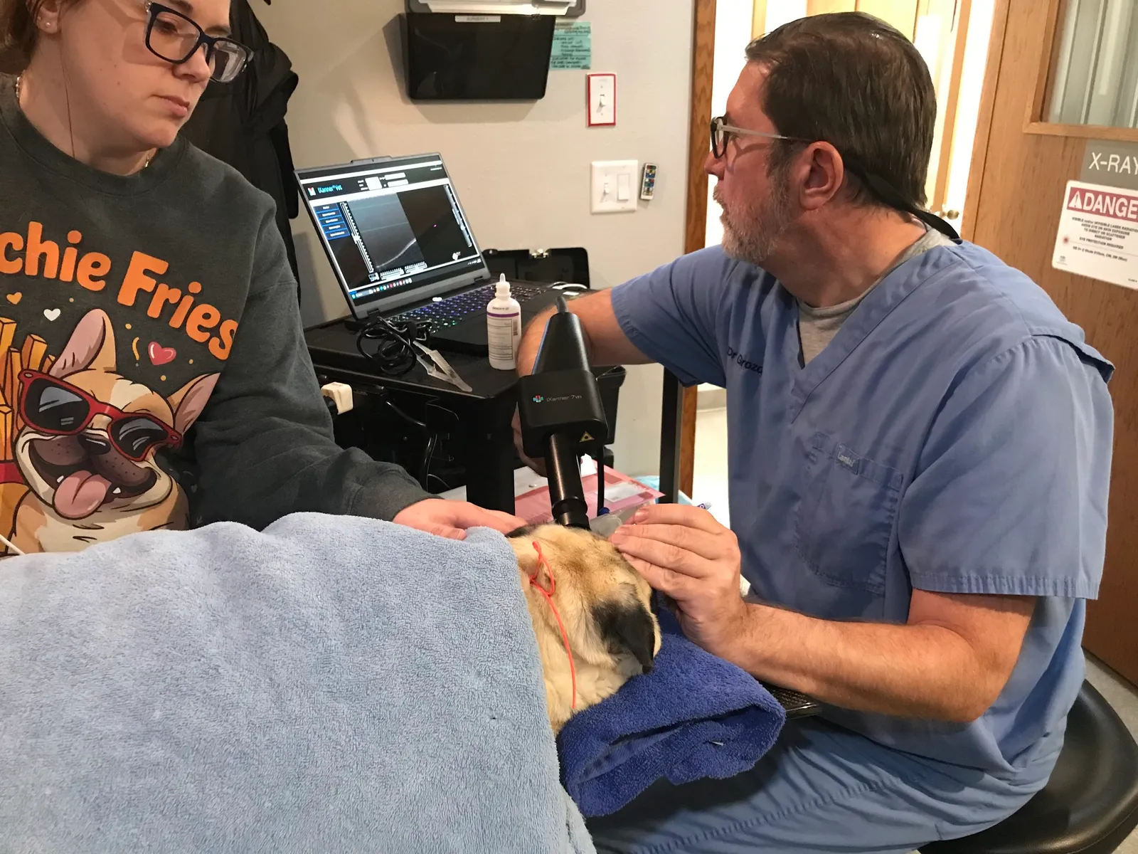
Handheld optical coherence tomography allows high-resolution imaging of the retina in veterinary patients.
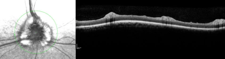
The fundus image identifies the scanned retinal region, while the OCT cross-section shows the layered structure of the retina in vivo.
Why OCT can be important
- Detects subtle retinal disease — OCT can identify microscopic retinal thinning, swelling, focal detachments, cystic change, photoreceptor disruption, and other abnormalities that may be difficult or impossible to see with fundoscopy alone
- Measures retinal structure objectively — Retinal thickness and individual retinal layers can be measured and compared over time, allowing us to monitor progression or response to treatment
- Helps localize the cause of vision loss — When combined with a complete ophthalmic examination, electroretinography (ERG), pupil testing, retinal photography, ultrasound, or advanced systemic testing, OCT can help distinguish retinal disease from optic nerve or central visual pathway disease
- Supports treatment planning and prognosis — Knowing whether retinal layers are preserved, inflamed, detached, or degenerating can influence treatment choices and help us discuss realistic expectations with pet owners
- Provides a permanent baseline — Images can be stored and repeated, making OCT useful for long-term follow-up of progressive retinal and optic nerve disorders
Conditions in which we may recommend OCT
- Sudden acquired retinal degeneration syndrome (SARDS) and other autoimmune or inflammatory retinopathies
- Cancer-associated retinopathy (CAR) and paraneoplastic retinal disease
- Progressive or hereditary retinal degeneration
- Retinal detachment, focal retinal separation, or subretinal fluid
- Retinal edema, cystic retinal change, or inflammatory retinal lesions
- Glaucoma and diseases affecting the optic nerve or retinal nerve fiber layer
- Unexplained vision loss when the retina appears relatively normal on routine examination
- Monitoring before and after selected medical, retinal, or vitreoretinal treatments
What happens during an OCT examination?
OCT is noninvasive and painless. A scanning camera is positioned close to the eye while a series of retinal images is obtained. In many cooperative dogs and cats, imaging can be performed while the patient is awake with gentle positioning. Some patients may benefit from light sedation, particularly if very precise scans are required or if they are anxious or unable to remain still.
The scan itself is rapid. The ophthalmologist then reviews the retinal layers, compares different retinal regions, evaluates both eyes, and interprets the images in the context of the complete eye examination and any other diagnostic testing.
OCT does not replace fundoscopy, ERG, or other ophthalmic tests. Instead, it adds a structural “microscope-like” view of the living retina that complements functional testing.
Intraoperative OCT: real-time imaging during surgery
Animal Eye Consultants of Iowa has intraoperative optical coherence tomography capability, allowing high-resolution OCT imaging to be obtained during advanced ophthalmic surgery. To our knowledge, we are the only veterinary institution in the world currently using intraoperative OCT in this way.
Unlike conventional OCT, which is performed before or after a procedure, intraoperative OCT provides cross-sectional structural information while the surgeon is operating. This can improve intraoperative surgical planning and execution by helping assess tissue configuration, retinal attachment, interfaces between the retina and vitreous, and the immediate anatomic effect of surgical maneuvers in real time.
For selected vitreoretinal and other advanced ophthalmic procedures, this additional information can help refine surgical decision-making without relying solely on the en face microscopic view.
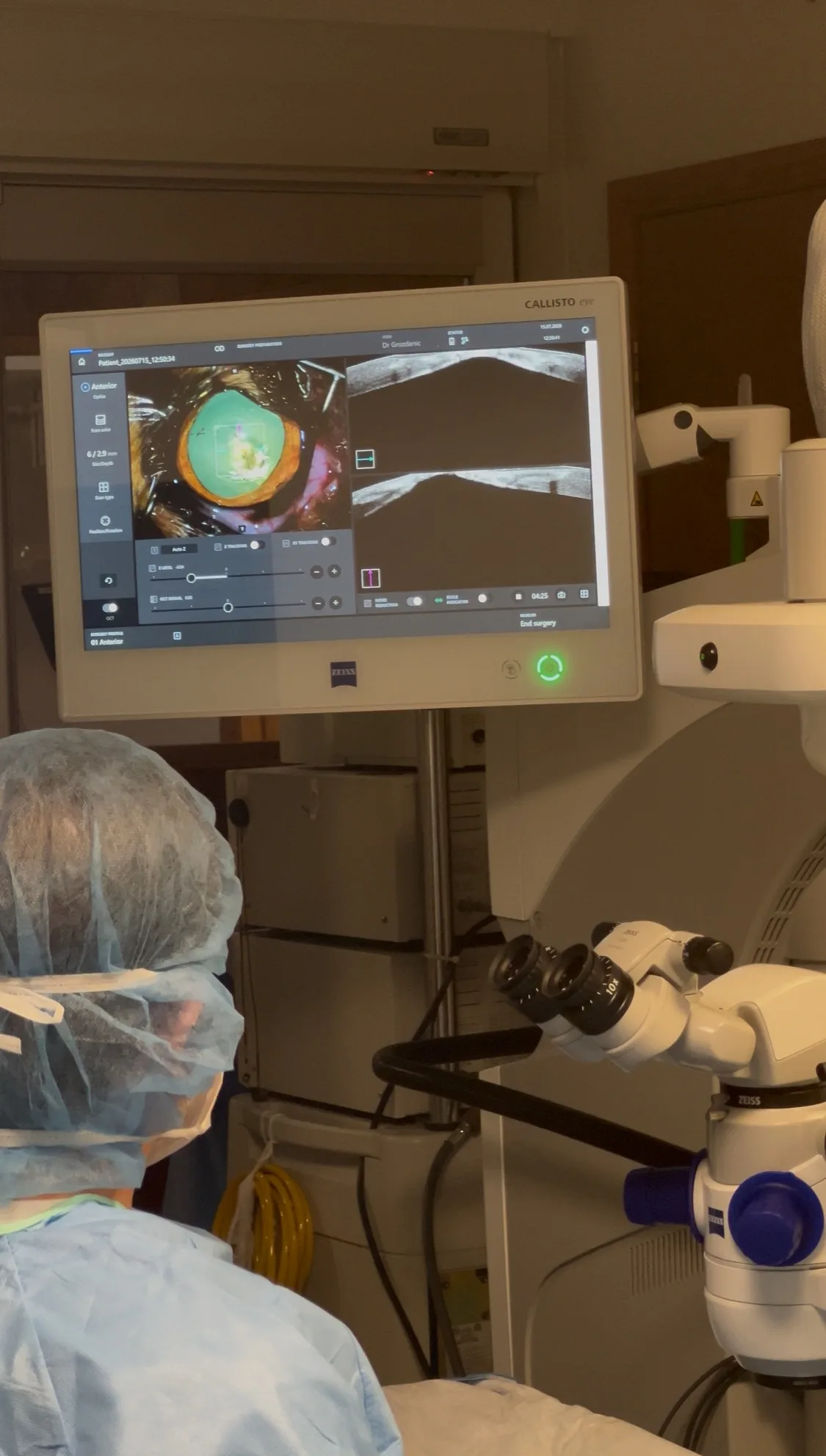
Intraoperative OCT integrated with the surgical microscope provides cross-sectional imaging during surgery, supporting real-time assessment of ocular structures and surgical maneuvers.
Our experience with veterinary OCT
Dr. Siniša D. Grozdanić has used optical coherence tomography in clinical and research studies of canine retinal disease, glaucoma, and comparative retinal anatomy for many years. His group published foundational Veterinary Ophthalmology studies defining normal spectral-domain OCT anatomy and retinal layer measurements in healthy dogs and characterizing normal mouse retinal structure and function with OCT. This normative work provides an important reference for recognizing disease-related structural abnormalities.
In a 2019 Veterinary Ophthalmology study of dogs with SARDS, Dr. Grozdanić and colleagues used OCT together with fundus photography, ERG, chromatic pupil testing, histology, and molecular analysis. OCT detected very small retinal detachments in 38% of the evaluated SARDS patients; these detachments were less than 1 mm wide and were not detectable by funduscopy or retinal photography. The study highlighted the ability of OCT to identify clinically important microscopic structural changes in canine retinal disease.
Earlier glaucoma research by Dr. Grozdanić and colleagues used OCT to measure retinal thinning after acute intraocular pressure elevation, illustrating the value of OCT for objective assessment of retinal structural injury. His later work on presumed cancer-associated retinopathy also incorporated OCT to identify photoreceptor disruption, focal retinal degeneration, retinal edema, and exudative detachments that helped characterize the disease process.
Frequently asked questions
Does OCT hurt?
No. OCT is a non-contact or near-contact optical imaging test and does not involve surgery, needles, or radiation.
Will my pet need anesthesia?
Usually not. Many patients can be imaged awake. Light sedation may occasionally be helpful when precise positioning is difficult.
How long does it take?
The scan itself is usually quick, although the total appointment time depends on the complexity of the eye examination and whether additional testing such as ERG, ocular ultrasound, or photography is needed.
Can OCT diagnose the cause of blindness by itself?
Not always. OCT shows retinal structure. The most accurate diagnosis often comes from combining OCT with the history, complete ophthalmic examination, ERG, pupil testing, laboratory testing, and other imaging when appropriate.
Can OCT be repeated?
Yes. Repeated scans are useful for tracking retinal thickness, lesions, detachments, or treatment response over time.
Scientific references
Presumed cancer-associated retinopathy mimicking SARDS in canines
Grozdanić SD, Lazić T, Kecova H, Mohan K, Adamus G, Kuehn MH. Veterinary Ophthalmology. 2021;24(2):125-155. doi:10.1111/vop.12853.
Optical coherence tomography and molecular analysis of SARDS eyes suggests the immune-mediated nature of retinal damage
Grozdanić SD, Lazić T, Kecova H, Mohan K, Kuehn MH. Veterinary Ophthalmology. 2019;22(3):305-327. doi:10.1111/vop.12597.
Spectral domain optical coherence tomography (SD-OCT) assessment of the healthy female canine retina and optic nerve
Hernandez-Merino E, Kecova H, Jacobson SJ, Hamouche KN, Nzokwe RN, Grozdanić SD. Veterinary Ophthalmology. 2011;14(6):400-405. doi:10.1111/j.1463-5224.2011.00887.x.
Characterization of structure and function of the mouse retina using pattern electroretinography, pupil light reflex, and optical coherence tomography
Mohan K, Harper MM, Kecova H, Ye EA, Lazić T, Sakaguchi DS, Kardon RH, Grozdanić SD. Veterinary Ophthalmology. 2012;15(Suppl 2):94-104. doi:10.1111/j.1463-5224.2012.01034.x.
Recovery of canine retina and optic nerve function after acute elevation of intraocular pressure: implications for canine glaucoma treatment
Grozdanić SD, Matić M, Betts DM, Sakaguchi DS, Kardon RH. Veterinary Ophthalmology. 2007;10(Suppl 1):101-107. doi:10.1111/j.1463-5224.2007.00584.x.
Concerned about unexplained vision loss or retinal disease?
A veterinary ophthalmic examination can determine whether OCT or other advanced retinal testing is appropriate for your pet.