
Ocular Ultrasound in Dogs and Cats
Ocular ultrasonography (eye ultrasound) is a rapid, noninvasive imaging technique used to evaluate structures inside the eye and orbit when they cannot be seen adequately during a routine ophthalmic examination. It is especially valuable when the cornea, anterior chamber, or lens is opaque—for example because of a dense cataract, corneal edema, hemorrhage, or severe inflammation.
What does ocular ultrasound show?
Using high-frequency sound waves, ultrasound creates real-time cross-sectional images of the globe, vitreous, retina, posterior segment, and tissues behind the eye. The examination can help determine whether the retina is attached, identify vitreal abnormalities or intraocular masses, detect blood or other material within the eye, evaluate orbital disease, and locate some foreign bodies.
Why this matters before treatment or surgery
When the back of the eye cannot be visualized directly, ultrasound can provide critical information about retinal integrity and other posterior-segment disease. These findings may influence prognosis, surgical planning, additional diagnostic testing, or whether referral for advanced ocular or orbital imaging is recommended.
How ocular ultrasound is performed
In most cooperative dogs and cats, ocular ultrasound can be performed awake with gentle restraint. A coupling gel is placed on the closed eyelid or, when appropriate, the examination is performed with a probe positioned at the ocular surface using standard ophthalmic technique. Images are obtained in multiple orientations so the ophthalmologist can evaluate the eye in real time.
The study is painless and usually takes only a few minutes. Sedation may be helpful in anxious, painful, or poorly cooperative patients. The need for sedation depends on the individual patient and the clinical situation.
Common reasons we recommend ocular ultrasound
- Dense cataract or other opacity that prevents examination of the retina and vitreous
- Suspected retinal detachment
- Vitreal degeneration, hemorrhage, inflammatory debris, or other posterior-segment abnormalities
- Suspected intraocular tumor or mass
- Trauma and possible intraocular foreign body
- Orbital swelling, pain, exophthalmos, suspected abscess, cyst, mass, or other retrobulbar disease
- Preoperative evaluation when visualization of the posterior segment is limited
Clinical examples
The following ultrasound images are taken from canine patients. The same ultrasound appearance can sometimes occur with more than one disease, so findings are interpreted together with the pet’s history and complete eye examination.
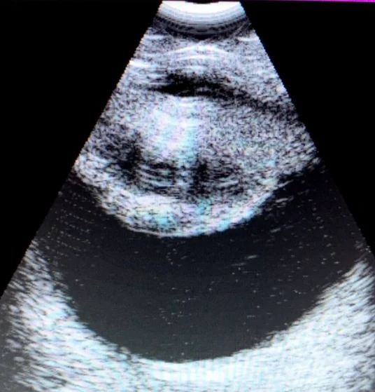
Cataract with normal-appearing vitreous and retina — a dense cataract may prevent direct visualization of the posterior segment. Here, ultrasound demonstrates a cataract while the vitreous cavity and posterior wall have a relatively normal appearance, useful information when assessing the posterior segment before surgical planning.
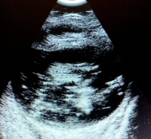
Severe vitreal degeneration — the normal vitreous is largely anechoic, meaning it appears dark on ultrasound. With advanced degeneration, mobile strands, membranes, or echogenic material may become visible. Ultrasound helps characterize these changes and distinguish them from other causes of abnormal echoes within the posterior segment.
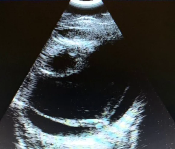
Cataract with retinal detachment — retinal detachment may be hidden behind a cataract or other opacity. On ultrasound, a detached retina often appears as a bright membranous structure separated from the posterior wall of the globe. Identifying it is important because it can substantially affect visual prognosis and may alter the recommended treatment plan.
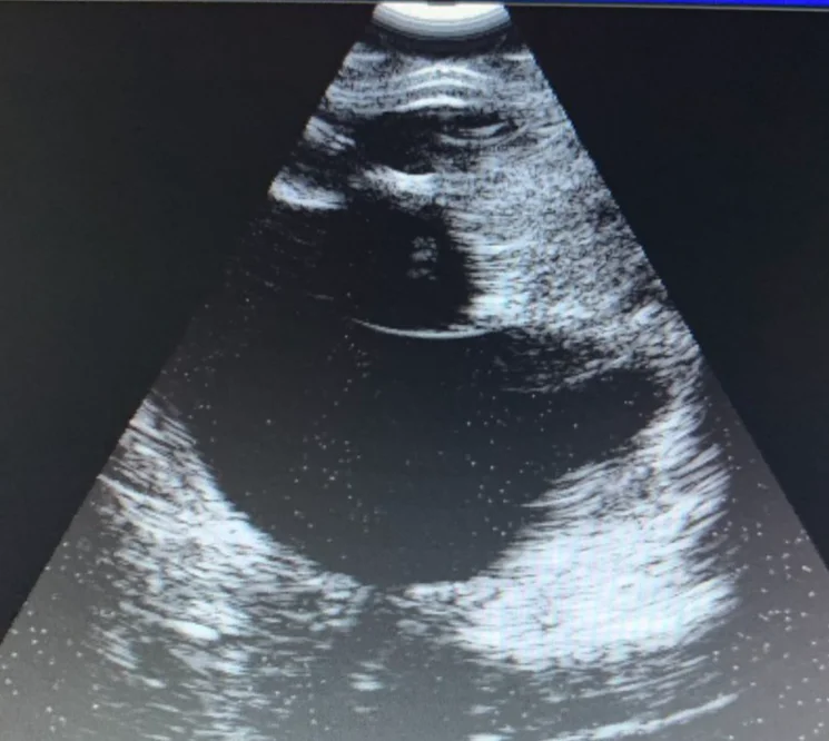
Intraocular tumor — ultrasound can help identify the size, shape, location, and internal characteristics of an intraocular mass, particularly when direct visualization is limited. It is also useful for assessing the relationship of a mass to the lens, vitreous, posterior wall, and other ocular structures.
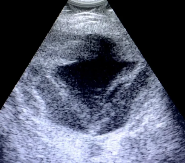
Retinal detachment with intraocular and subretinal blood — blood within the eye can obscure normal anatomy and create complex ultrasound echoes. Ultrasound is especially helpful in hemorrhagic eyes because it allows evaluation of structures that cannot be seen through the blood.
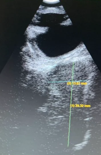
Measurement of a posterior-segment abnormality — ultrasound allows electronic measurements directly on the image. Depending on the case, measurements can document lesion size, globe dimensions, the extent of a mass or fluid collection, or changes over time during follow-up examinations.
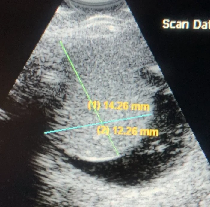
Large intraocular tumor — large masses can distort normal ocular anatomy. Ultrasound provides a rapid way to document the dimensions and internal echogenicity of a lesion. Depending on the suspected diagnosis, further imaging such as CT or MRI and systemic staging may be recommended.
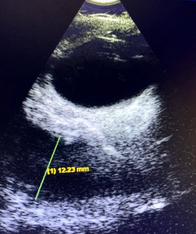
Orbital abscess — ultrasound can extend beyond the globe to evaluate the orbit. Fluid-filled or mixed echogenic lesions behind the eye may be detected and measured. In a patient with orbital pain, swelling, or protrusion of the eye, ultrasound can help identify an abscess or other space-occupying lesion and guide the next diagnostic steps.
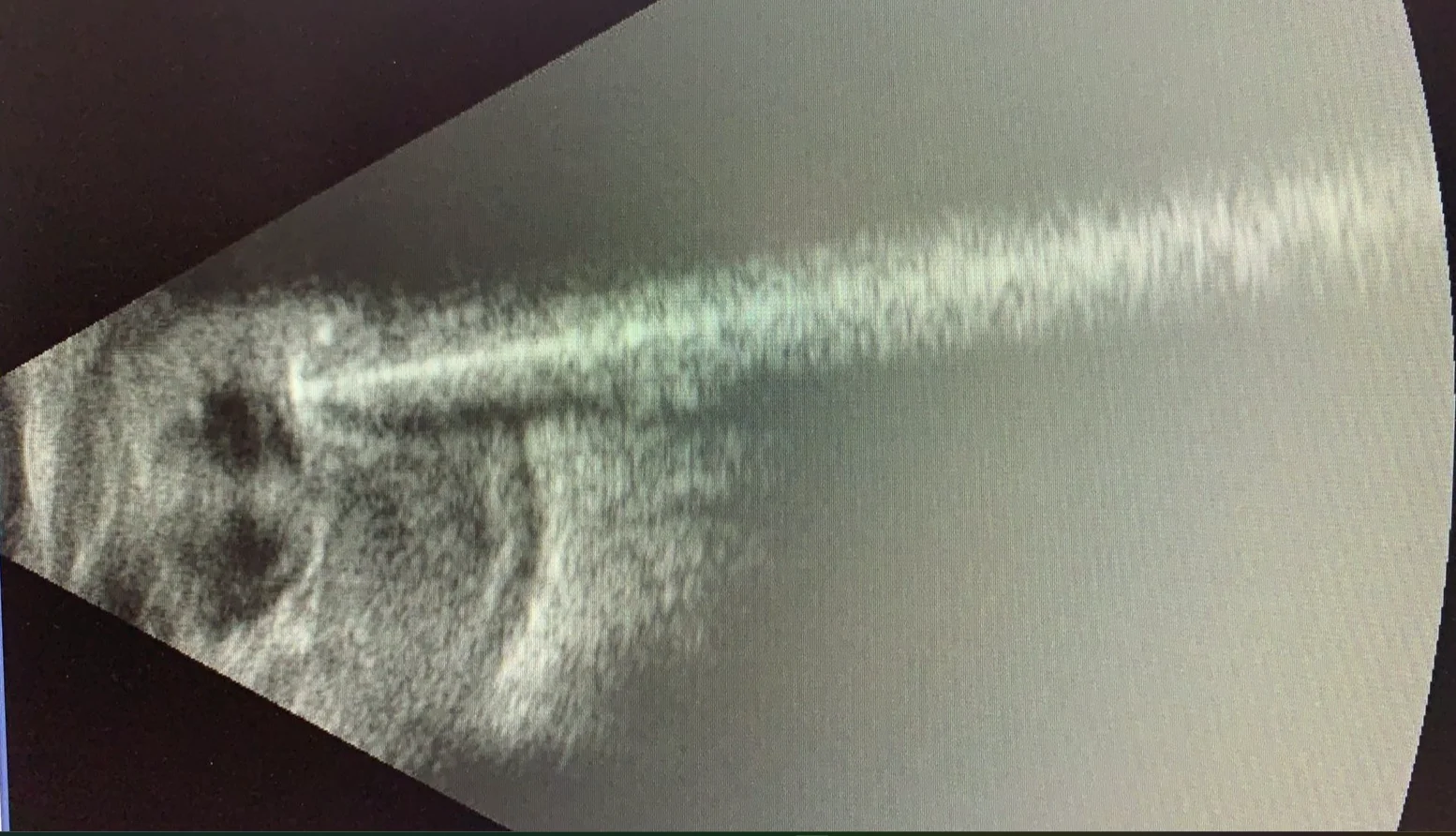
Intraocular metallic foreign body (gun pellet) — metallic material is strongly reflective on ultrasound and can produce characteristic reverberation or comet-tail artifacts. In traumatic cases, ultrasound may help localize a foreign body and evaluate associated intraocular damage. When a penetrating injury is suspected, the technique is modified to avoid pressure on the globe, and complementary imaging such as CT may also be indicated.
What ocular ultrasound can and cannot tell us
Ocular ultrasound is an imaging test that complements, rather than replaces, a complete veterinary ophthalmic examination. It is very useful for structural assessment when direct visualization is limited, but the ultrasound appearance of a lesion is not always specific for one diagnosis. Clinical history, slit-lamp examination, intraocular pressure measurement, fundus examination when possible, laboratory testing, cytology or biopsy, and CT/MRI may still be needed.
Important limitation
Ultrasound findings must be interpreted together with the complete eye examination. Similar ultrasound appearances can be produced by different diseases, and some very small or superficial lesions may be better assessed by other ophthalmic imaging techniques.
Frequently asked questions
Does ocular ultrasound hurt?
The examination is noninvasive and is generally well tolerated. Most patients require only gentle restraint. Sedation is considered when needed for comfort and safety.
Can ultrasound be performed when a dog or cat has a cataract?
Yes. A cataract is one of the most common reasons to perform ocular ultrasound because the opaque lens may block direct examination of the retina and vitreous.
Can ultrasound diagnose a retinal detachment?
Ultrasound is one of the most useful tests for detecting retinal detachment when the retina cannot be seen directly. The appearance and extent of the detachment are interpreted by the ophthalmologist in the context of the complete examination.
Can ultrasound detect tumors behind or inside the eye?
It can identify many intraocular and orbital masses, document their location and dimensions, and help determine whether additional imaging or diagnostic sampling is appropriate.
Is CT or MRI sometimes still necessary?
Yes. CT or MRI may provide additional information about the orbit, skull, optic nerve, adjacent tissues, or suspected foreign material, and may be recommended depending on the clinical findings.
When your pet may benefit from ocular ultrasound
If your pet has a dense cataract, sudden vision loss, suspected retinal disease, ocular trauma, an intraocular mass, or signs of orbital disease, a veterinary ophthalmic examination can determine whether ocular ultrasound would provide useful additional information. Contact Animal Eye Consultants of Iowa to discuss evaluation and imaging options for your pet.
Educational images on this page illustrate representative ultrasound findings from canine patients. Individual patients may look different.