
Fundoscopy in Dogs and Cats
Fundoscopy is the examination of the back of the eye—the retina, optic nerve, retinal blood vessels and underlying choroid. It is one of the most important parts of a complete eye examination because many retinal diseases can threaten vision before changes are obvious from the outside.
What is fundoscopy?
The fundus is the inside surface at the back of the eye. During fundoscopy, a veterinary ophthalmologist directs light through the pupil and examines the retina and optic nerve using an ophthalmoscope or specialized imaging system. The examination helps determine whether the retina is healthy, inflamed, degenerating, detached, bleeding or affected by a systemic disease.
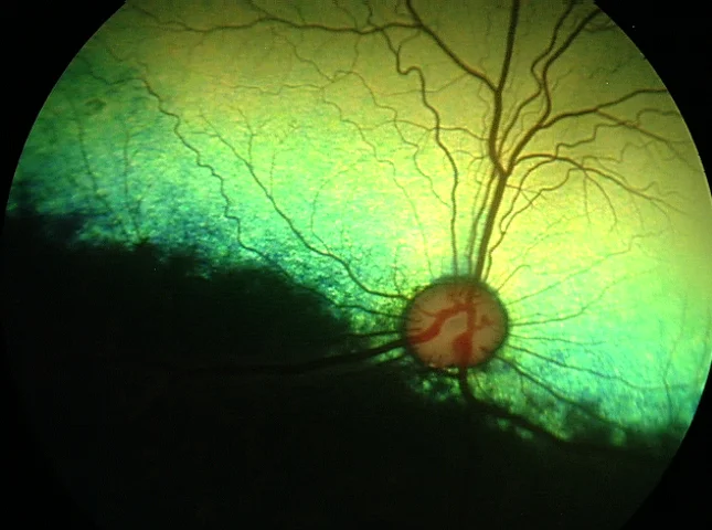
Normal canine fundus. The reflective tapetum, optic nerve and retinal blood vessels can be examined directly during fundoscopy.
Retina
The retina contains the light-sensitive cells that convert light into signals sent to the brain. Retinal degeneration, inflammation and detachment can all affect vision.
Optic nerve
The optic nerve carries visual information from the eye to the brain. Changes in its size, color or contour may indicate developmental, inflammatory or degenerative disease.
Blood vessels & choroid
Retinal vessels and the choroid can show evidence of hypertension, inflammation, infection, hemorrhage, vascular congestion and other systemic disorders.
What should you expect during the examination?
Fundoscopy is generally quick and non-invasive. Most dogs and cats do not require sedation. In many patients, dilating drops are used so the veterinarian can examine a larger portion of the retina.
- Initial eye exam — The front of the eye, pupils, vision responses and eye pressure are assessed first.
- Pupil dilation — Dilating drops may be placed in the eye to improve visualization of the retina and optic nerve.
- Fundus examination — The veterinarian examines the retina with a bright light and magnifying optical system.
- Additional testing — Depending on the findings, retinal photography, ocular ultrasound, ERG, blood pressure measurement or laboratory testing may be recommended.
Normal appearance varies between patients
A normal fundus does not look identical in every dog or cat. Coat color, iris pigmentation, breed, age and development of the tapetum can all influence appearance. Recognizing these normal variations is essential before deciding that a retinal change is abnormal.
Typical tapetal and non-tapetal fundus — a well-developed green-yellow reflective tapetum superiorly and a darker pigmented non-tapetal region inferiorly, with retinal blood vessels radiating from the optic nerve.
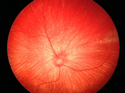
Subalbinotic canine fundus — dogs with reduced pigmentation may have a reddish fundus with prominent visible choroidal vessels, a normal variation commonly seen in lightly pigmented animals.
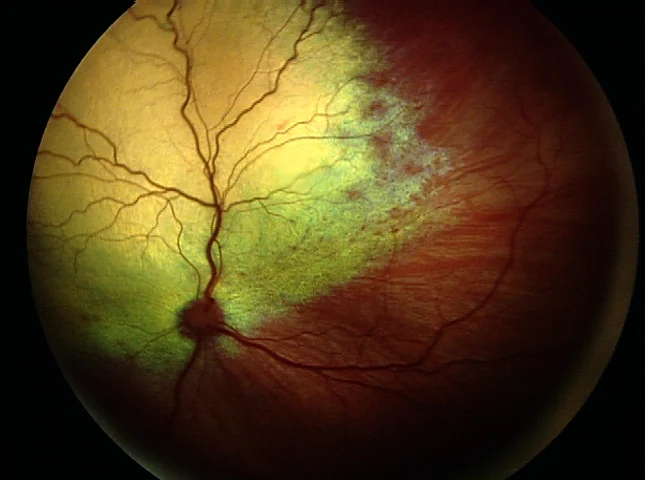
Normal feline fundus — the feline optic nerve is normally smaller and darker than the canine optic nerve because it lacks the same degree of visible myelination, and pigmentation can vary substantially between cats.
Examples of diseases detected during fundoscopy
The following examples are taken from veterinary teaching cases. The same visual pattern can sometimes occur with more than one disease, so fundus findings are interpreted together with the pet’s history, complete eye examination and additional diagnostic testing.
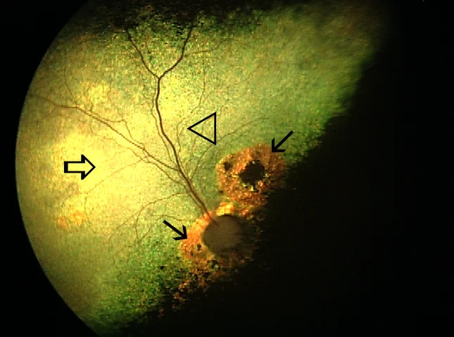
Progressive retinal atrophy (PRA) — an inherited retinal degeneration in many breeds. Typical fundus changes include increased reflectivity of the tapetum and progressive narrowing of retinal blood vessels. Night vision is often affected before day vision.
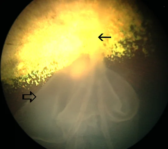
Complete retinal detachment — a detached retina is separated from the tissues that normally support it. Detachment can cause sudden or progressive visual impairment and may occur with retinal tears, inflammation, hypertension, infection or other ocular disease.
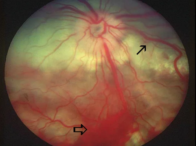
Hypertensive retinopathy in a dog — high systemic blood pressure can damage retinal vessels. Findings may include retinal hemorrhage, edema, vascular congestion and, in severe cases, retinal detachment.
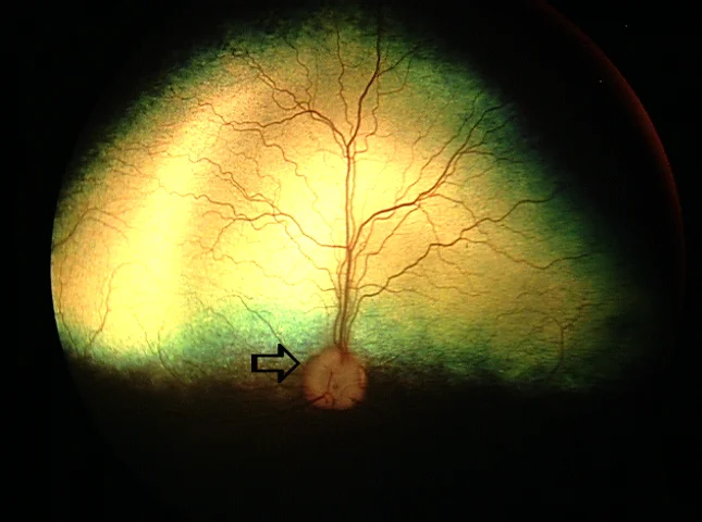
SARDS: severe vision loss with few early fundus changes — in sudden acquired retinal degeneration syndrome, the retina may appear relatively normal during the early stages despite profound vision loss. Electroretinography and other testing can therefore be important when the fundus does not explain the degree of blindness.
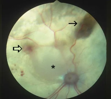
Hypertensive retinal disease in a cat — systemic hypertension is an important cause of acute visual loss in cats. Fundoscopy may reveal hemorrhage, choroidal or retinal fluid accumulation and bullous retinal detachment. Blood pressure measurement is especially important in older cats and cats with kidney or thyroid disease.
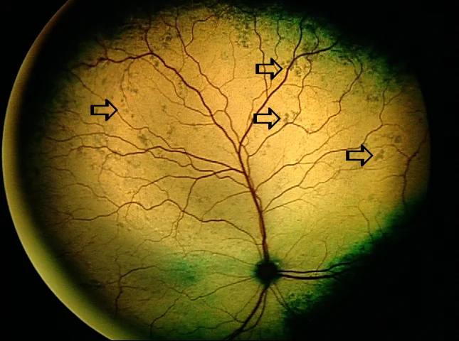
Chorioretinal inflammation — inflammatory or infectious disease may produce focal or multifocal lesions in the retina and choroid. This teaching example shows choroidal exudative lesions associated with toxoplasmosis.
Important
A fundus photograph is only one part of the diagnostic process. Similar retinal appearances can be caused by different diseases, and some serious retinal disorders may produce very few visible changes early in their course.
When is fundoscopy especially important?
- Sudden or gradual vision loss
- Difficulty seeing at night or in dim light
- Dilated pupils or abnormal pupil responses
- Reluctance to jump, use stairs or navigate unfamiliar areas
- Suspected retinal detachment or retinal degeneration
- Systemic hypertension
- Kidney disease, hyperthyroidism or other diseases associated with hypertension
- Suspected infectious, inflammatory or immune-mediated disease
- Breed-related screening for inherited eye disease
- Preoperative or postoperative assessment when retinal health is important
What if the retina cannot be seen?
If cataract, corneal opacity, bleeding or another problem blocks the view of the back of the eye, fundoscopy may not be possible. In those patients, ocular ultrasound can help evaluate the retina and vitreous, while electroretinography (ERG) can assess retinal function in selected cases.
These tests are complementary: fundoscopy shows the appearance of the retina, ultrasound shows its structure when the view is blocked, and ERG evaluates retinal electrical function.
Concerned about your pet's vision?
Changes such as bumping into objects, missing treats or toys, hesitation in dim light, dilated pupils or sudden blindness should be evaluated promptly. A complete veterinary ophthalmic examination—including fundoscopy when possible—can help determine whether the problem arises from the retina, optic nerve, other parts of the eye or the nervous system.
Educational images on this page illustrate representative fundus appearances from veterinary teaching cases. Individual patients may look different.