
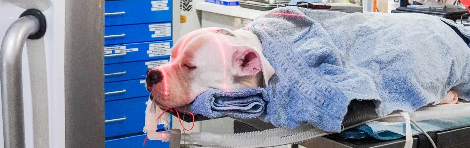
CT Imaging for Veterinary Ophthalmology
Advanced imaging of the orbit, skull, nasolacrimal system and periocular tissues, with CT available for diagnostic evaluation, surgical planning and selected intraoperative applications.
CT Imaging at Animal Eye Consultants of Iowa
Computed tomography (CT) provides cross-sectional and three-dimensional visualization of structures that can be difficult or impossible to evaluate completely during a routine ophthalmic examination. At Animal Eye Consultants of Iowa, CT is integrated with specialized ophthalmic examination, ocular and orbital ultrasonography, electrodiagnostics, endoscopy, and surgery when needed. This is particularly valuable for disorders involving the orbit, skull, paranasal structures, optic nerve region, nasolacrimal system, and periocular tissues.
When CT may be recommended
- Orbital disease, including suspected tumors, abscesses, cellulitis, inflammatory disease, hemorrhage, and foreign bodies
- Exophthalmos, enophthalmos, painful or restricted globe retropulsion, periocular swelling, or protrusion of the third eyelid
- Unexplained visual loss when disease behind the globe, at the optic nerve, or within the skull is suspected
- Nasolacrimal obstruction or disease involving adjacent nasal and facial structures
- Preoperative mapping of orbital and periocular lesions and planning of surgical access
- Selected intraoperative procedures in which imaging can confirm anatomy, implant position, bleeding, or other changes during surgery
Cone-beam CT and ophthalmic imaging
Cone-beam CT (CBCT) systems are compact and can be positioned close to the surgical field. CBCT provides high-contrast imaging of head and orbital anatomy and can be used for intraoperative imaging. Compared with conventional multi-slice CT, CBCT generally offers lower radiation exposure and a smaller, mobile footprint, while conventional CT provides superior soft-tissue contrast in many body regions. The optimal imaging modality depends on the clinical question.
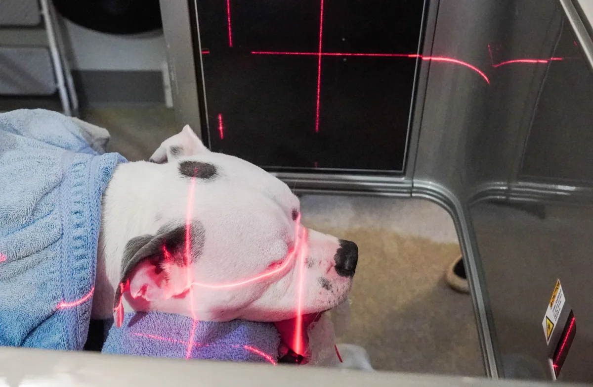
Laser alignment ensures accurate positioning of the head.
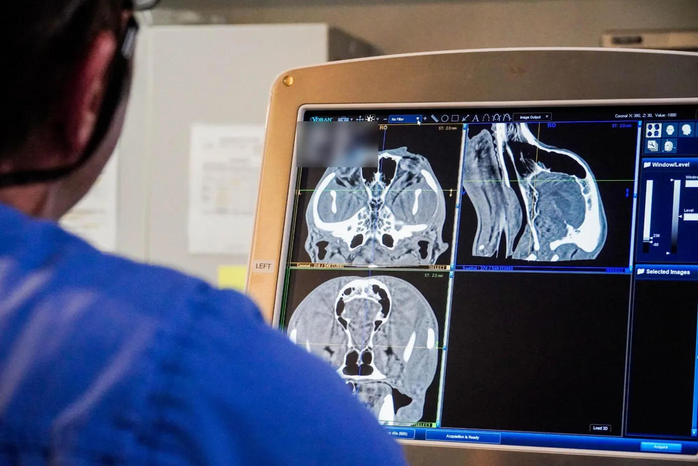
CT images are reviewed in multiple planes on the scanner workstation.
Clinical examples
The following cases illustrate how CT can change diagnosis, treatment planning, and surgical decision-making in veterinary ophthalmic patients.
Case example 1 – Deep orbital foreign body
A young Labrador Retriever had intermittent mucopurulent discharge from the right eye despite a largely normal ophthalmic examination and normal nasolacrimal flushing. Limited third-eyelid mobility prompted advanced imaging.
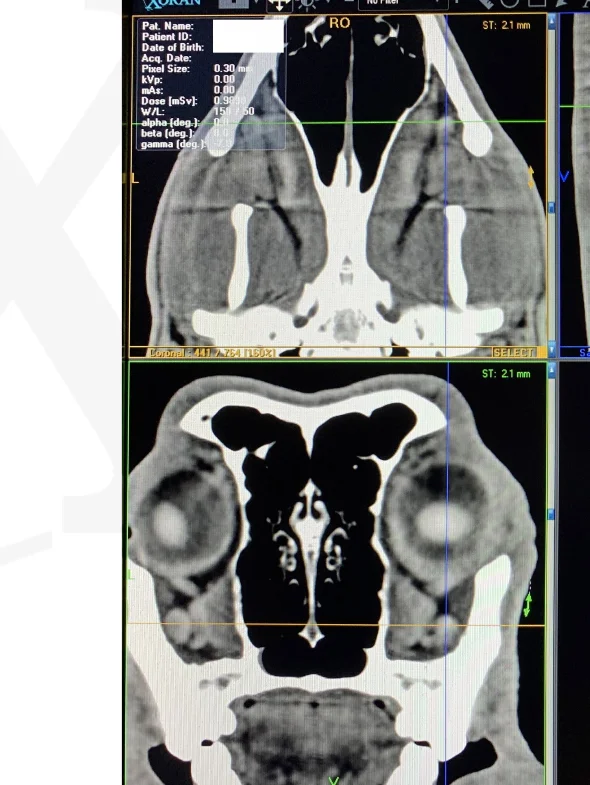
CT finding: CT localized a deep orbital abnormality that was subsequently confirmed surgically as a wooden foreign body.
Clinical value: Localization before surgery can help determine the safest approach to a deep orbital lesion and can reveal disease that is not apparent on the ocular surface.
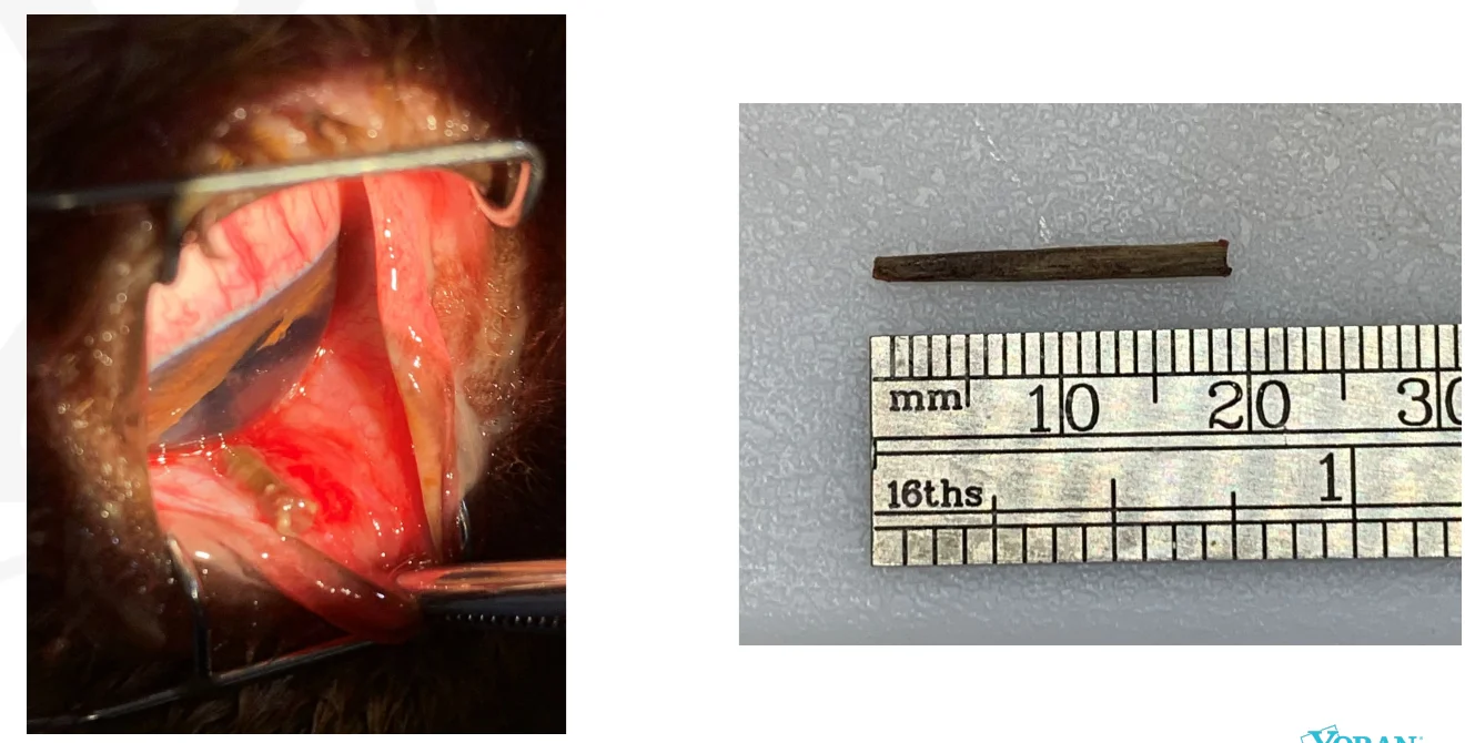
Surgical removal of the wooden orbital foreign body.
Case example 2 – Orbital mass
An adult cat developed slowly progressive swelling around one eye, third-eyelid protrusion, exophthalmos, and reduced retropulsion without marked pain.
CT finding: CT demonstrated an orbital mass and defined its location and extent relative to the globe and surrounding structures.
Clinical value: Cross-sectional imaging helps guide biopsy, endoscopic evaluation, surgical planning, and discussion of prognosis when an orbital mass is suspected.
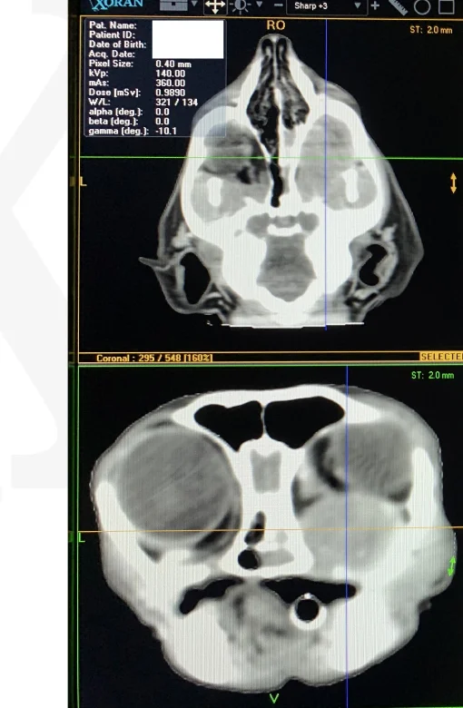
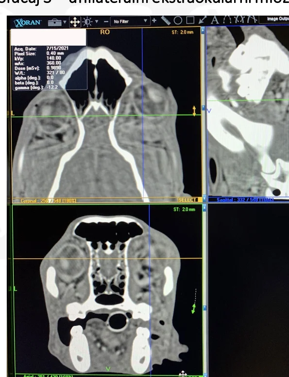
Case example 3 – Extraocular myositis with compressive optic neuropathy
A young Labrador Retriever developed rapidly progressive periocular swelling, painful exophthalmos, and acute blindness despite a normal-appearing retina and optic nerve on intraocular examination.
CT finding: CT showed enlargement of the extraocular muscles consistent with severe unilateral extraocular myositis and compression in the orbital apex/optic nerve region.
Clinical value: The imaging findings supported urgent orbital decompression and medical treatment. The case documents recovery of vision and pupillary/photopic responses after treatment.
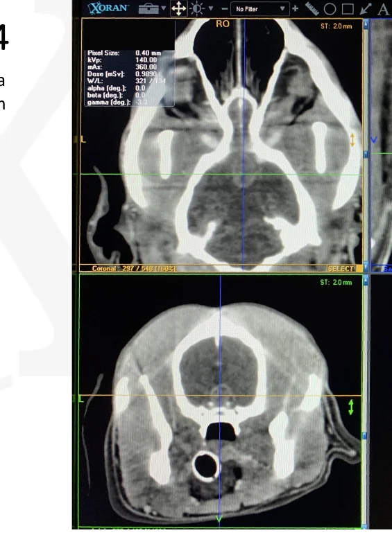
Case example 4 – Intracranial disease associated with ocular signs
A Labrador Retriever with progressive visual dysfunction, vitritis, and exudative chorioretinal lesions underwent CT after routine systemic testing did not identify a cause.
CT finding: CT demonstrated a pituitary-region mass, associated clinically with paraneoplastic vitritis and chorioretinitis.
Clinical value: Advanced imaging can be valuable when ocular findings suggest disease beyond the eye itself, especially when neurologic, optic-nerve, or intracranial involvement is possible.
Case example 5 – Orbital abscess and compressive optic neuropathy
A Chihuahua developed severe painful periocular swelling and visual loss shortly after a dental procedure. The patient was too painful for a complete orbital ultrasound without general anesthesia.
CT finding: CT identified an orbital abscess with compression of the optic nerve region.
Clinical value: Imaging supported urgent surgical drainage and decompression. In this case, vision and pupillary/photopic responses recovered within days after surgery and treatment.
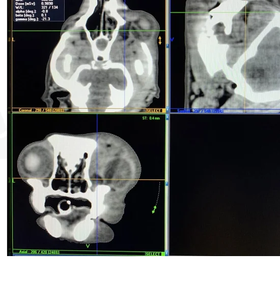
Case example 6 – Panophthalmitis, cellulitis and secondary glaucoma
A Doberman with progressive periocular swelling, corneal ulceration, glaucoma, and blindness underwent CT to evaluate the full extent of orbital and ocular disease.
CT finding: CT demonstrated extensive inflammatory disease compatible with panophthalmitis and cellulitis with secondary glaucoma.
Clinical value: CT can define how far infection or inflammation extends beyond the globe and can support decisions regarding sampling, surgery, and systemic investigation.
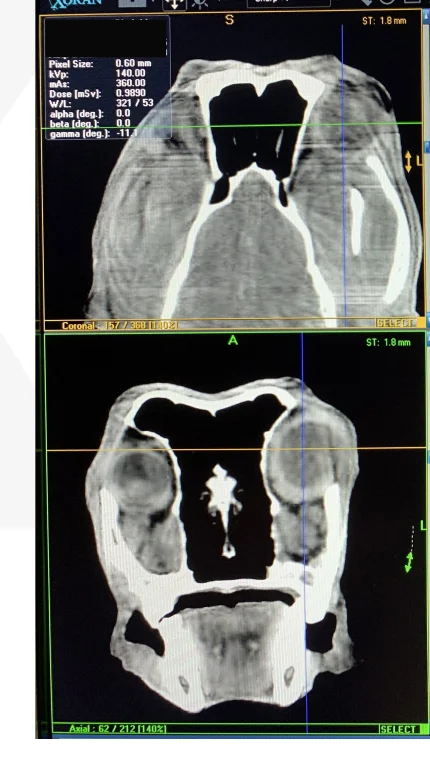
Intraoperative CT: imaging during surgery
One of the most distinctive applications of the CT system is the ability to acquire images during selected orbital procedures. Intraoperative imaging can confirm anatomy and surgical progress without relying only on external appearance or palpation. In one case, CT was used during orbital implant surgery when acute swelling developed before the end of the procedure.
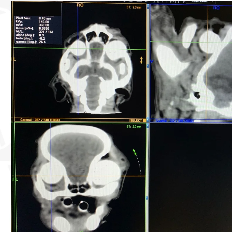
Intraoperative CT demonstrated accumulation of blood between the orbital fascia and skin, allowing immediate treatment.
CT demonstrated blood accumulation between the orbital fascia and skin. Aspiration and a compression bandage were then performed. This illustrates how intraoperative CT can provide immediate anatomic information when a surgical complication is suspected.
What owners and referring veterinarians can expect
- A complete ophthalmic examination is performed first to determine whether CT is the most appropriate imaging test.
- Sedation or general anesthesia may be required so that the head remains accurately positioned and the patient remains comfortable and still.
- Depending on the clinical question, CT may be combined with contrast administration, orbital ultrasound, endoscopy, sampling, biopsy, or surgery.
- Images are interpreted together with the ophthalmic examination and other diagnostic findings to develop a targeted treatment plan.
Referral indications
Consider referral for CT evaluation when a patient has unexplained exophthalmos or enophthalmos, orbital pain, periocular swelling, restricted globe movement or retropulsion, third-eyelid protrusion, suspected orbital mass or foreign body, recurrent orbital inflammation, suspected nasolacrimal disease, acute visual loss with a normal-appearing intraocular examination, or when preoperative orbital mapping would improve surgical planning.
Scientific references
Preoperative and intraoperative CT imaging for orbital foreign bodies identification and surgical planning in veterinary medicine
Grozdanić SD, Murtha H, Lazić T, Đukić S, Luzetskii S, Ursu DC, Sarment D. Acta Veterinaria-Beograd. 2024;74(3):367-397. doi:10.2478/acve-2024-0026.
- The study had three parts: a canine skull model with nine masked scenarios using metal, wood, plastic and glass objects, a test of a surgical navigation system, and three clinical dogs treated between 2020 and 2023.
- Metal and glass were detected best, wood was variable, and plastic was moderate to low. Porcupine quills were found more reliably with ultrasound than with CT.
Successful medical treatment of an orbital osteoma in a dog
Grozdanić S, Riedesel EA, Ackermann MR. Veterinary Ophthalmology. 2013;16(2):135-139. doi:10.1111/j.1463-5224.2012.01029.x.
- CT showed a smooth bony mass in the orbit without bone destruction. After treatment with anti-inflammatory drugs (NSAIDs), the mass regressed completely, and the dog had no signs for 5 years.
Endoscopic diagnostic and surgical orbital approach in canines
Đurić ML, Krstić VP, Lazić TM, Grozdanić SD. Acta Veterinaria Hungarica. 2022;70(2):100-112. doi:10.1556/004.2022.00017.
- The paper describes several endoscopic routes into the orbit in cadavers and in clinical patients. The route through the dorsal orbital ligament was the hardest to perform but gave the best view of the orbit.
Talk with us about CT imaging
If your pet has unexplained eye or orbital changes, or you are a veterinarian considering referral, contact Animal Eye Consultants of Iowa to discuss whether CT is the right next step.
Educational images on this page are from individual patients. Findings in other patients may look different.