
Chromatic Pupil Light Reflex (cPLR) Testing
A rapid red- and blue-light test that helps detect retina and optic nerve disease.
A 30-second light test for your pet's vision
Many retina and optic nerve problems begin quietly. A pet may still appear to see normally while the cells at the back of the eye are already failing. Chromatic pupil light reflex (cPLR) testing uses carefully selected red and blue light to check how the pupil responds, giving our ophthalmology team fast, non-invasive information about the functional health of the retina and optic nerve.
The test is quick, painless, and performed in the exam room. In many patients, an abnormal pupil response to red and blue light is one of the earliest signs of retina or optic nerve disease and can appear months before obvious vision loss, which creates a window for prompt treatment.
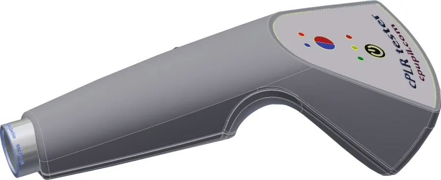
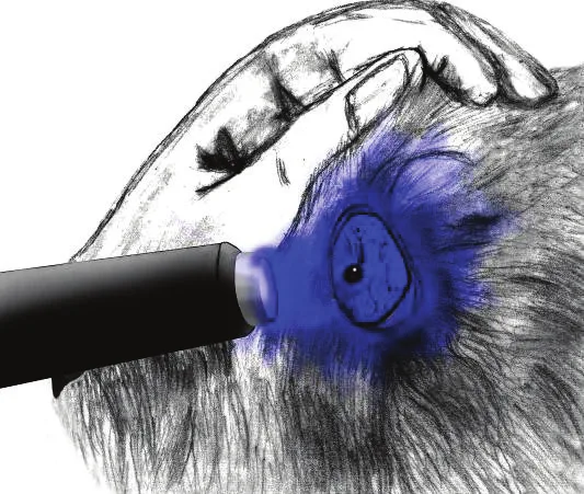
The handheld cPLR Tester delivers calibrated red and blue light stimuli.
What is chromatic pupil light reflex testing?
When light enters the eye, the pupil constricts. This pupil light reflex depends on healthy light-sensing cells in the retina, an intact optic nerve, and the brain pathways that control the pupil. cPLR testing separates these contributions by using two colors of light:
- Red light (about 630 nm) mainly tests the rods and cones, the photoreceptors that dogs rely on for everyday vision.
- Blue light (about 480 nm) also strongly activates melanopsin, a light-sensitive pigment found in a special group of retinal ganglion cells (intrinsically photosensitive retinal ganglion cells, or ipRGCs). These cells can drive a pupil response even when rods and cones are not working.
By comparing the pupil response to each color, the ophthalmologist can tell whether a problem is more likely located in the outer retina (photoreceptors) or in the optic nerve.
Why early testing matters
In many patients, abnormal pupil responses to chromatic stimuli are very early signs of retina or optic nerve disease and can precede vision problems by months, allowing immediate therapeutic intervention.
- Fast: the testing procedure can be completed in about 30 seconds per eye.
- Non-invasive: only light is used; nothing touches the eye.
- Informative: helps localize disease to the retina or optic nerve, and helps decide which additional tests are needed.
- Useful when the retina cannot be seen: in pets with cataracts, cPLR testing provides information about retina and optic nerve function before cataract surgery is considered.
What happens during the test
Your pet stays awake and comfortable. The room lights are dimmed for a short time so the pupils can relax and dilate. The ophthalmologist then holds the cPLR Tester about one inch from each eye and shines red light, followed by blue light, for about 10 seconds each while watching how much and how quickly the pupil constricts. Short rest periods in the dark between flashes allow the pupil to dilate again.
Red-light stimulation
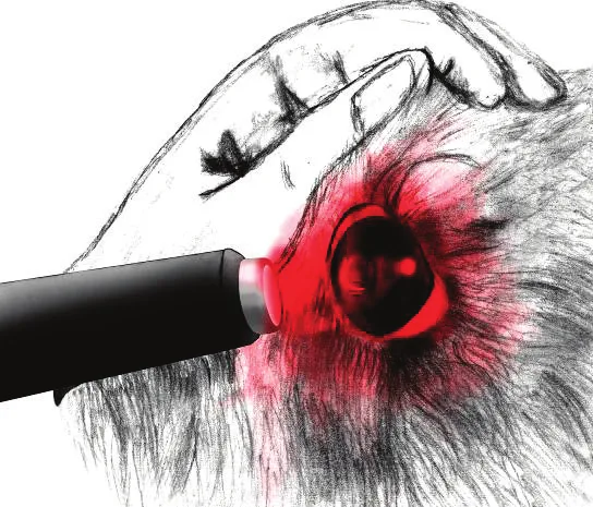
Start of the test: dilated pupil
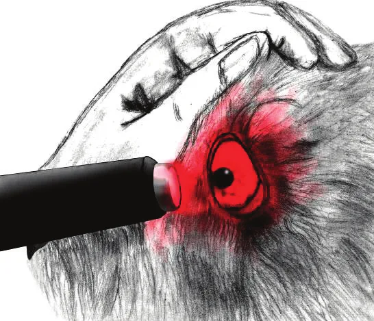
End of the test: constricted pupil
Blue-light stimulation
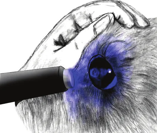
Start of the test: dilated pupil
End of the test: constricted pupil
In healthy eyes, strong light of either color produces rapid and complete pupil constriction. Blue light usually produces somewhat faster and more complete constriction.
What the results can tell us
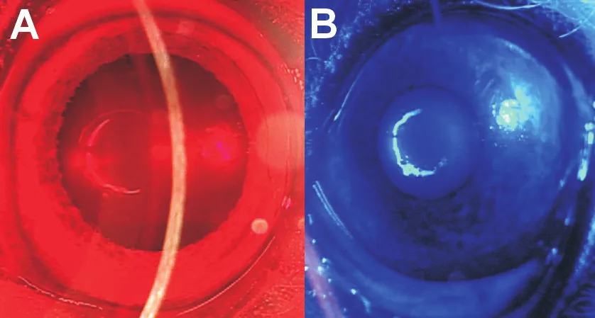
No red, good blue: retinal disease
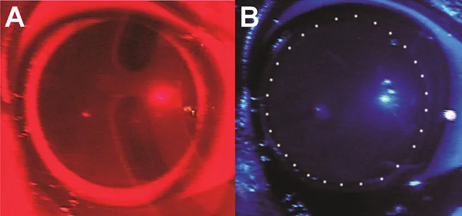
No red, no blue: optic nerve disease
Important: cPLR testing is a screening and localization tool. It does not establish a final diagnosis on its own. Results are interpreted together with your pet's history, a complete ophthalmic examination, and, when needed, electroretinography (ERG), optical coherence tomography (OCT), ultrasound, and laboratory testing.
The science behind the test
Different light colors activate different retinal cells
Blue light of 480 nm activates short and medium-long wavelength cone opsins, melanopsin, and rhodopsin. Red light of 630 nm cannot activate short-wavelength cone opsin or melanopsin. Because 630 nm red light activates only the far edge of the rhodopsin and medium-long wavelength cone sensitivity spectrum, disease in these photoreceptors (rods and medium-long wavelength cones are the dominant photoreceptors in the canine retina) can show up as an incomplete pupil light reflex, or as pupil “escape,” particularly after red light.
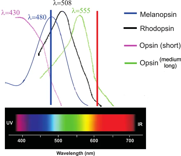
Spectral sensitivity of canine retinal photopigments. Image provided by Dr. Siniša D. Grozdanić.
Melanopsin and non-melanopsin pathways
Red light activates the rod–cone pathway. Disease in the rods, cones, or the inner retinal neurons that connect to ipRGCs reduces the pupil response to red light, while the melanopsin-driven response to bright blue light can be preserved.
Optic nerve lesions impair signal transmission from both melanopsin and non-melanopsin pathways, so the pupil response is reduced regardless of the light color used. In patients who recover from optic neuritis, damage to ipRGCs may prevent recovery of the pupil light reflex and dazzle response, yet vision can still be processed if enough other retinal ganglion cells remain.
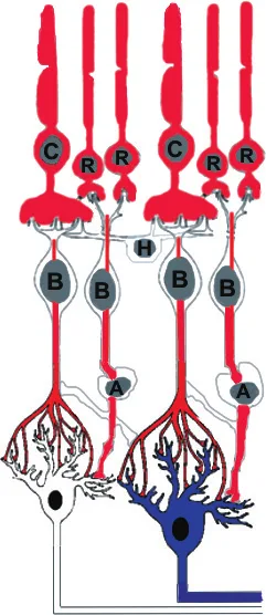
C = cone; R = rod; H = horizontal cell; B = bipolar cell; A = amacrine cell.
Conditions cPLR testing can help evaluate
- Sudden acquired retinal degeneration syndrome (SARDS) and other immune-mediated retinopathies, which typically show the “no red, good blue” pattern
- Inherited and acquired retinal degeneration, including progressive retinal atrophy
- Retinal detachment
- Optic neuritis and other optic nerve diseases
- Cataract patients, to screen retina and optic nerve function when the back of the eye cannot be examined directly
- Unexplained vision loss, dilated pupils, or night blindness
cPLR testing is used in both dogs and cats as part of a complete neuro-ophthalmic examination.
For referring veterinarians
cPLR testing in your hospital
cPLR testing is most effective when it is introduced as part of the regular physical examination of each patient. Detailed ocular examination is rarely performed during the annual wellness visit, but chromatic pupil light reflex testing allows rapid screening for possible retina and optic nerve abnormalities without interrupting a busy daily patient flow. Early detection allows timely intervention and can help prevent progression to a stage where treatment is very difficult and expensive.
Testing protocol with the cPLR Tester
- Turn on the switch on the back of the instrument.
- Press the power button. The instrument starts automatically in red-light mode (the red diode on the control panel lights). Turn off the examination room light and dark-adapt the patient for 10 seconds.
- Position the instrument one inch in front of the eye and activate it by pressing the side button.
- Illuminate the left eye with red light from one inch for 10 seconds and observe the change in pupil diameter. Remove the light after 10 seconds or when the pupil is maximally constricted (usually 3–4 seconds).
- Wait 30 seconds for dark adaptation and full pupil dilation, then illuminate the right eye with red light for 10 seconds and observe the change in pupil diameter.
- Press the red-blue button to switch to blue-light mode (the blue diode lights) and wait 30 seconds for pupil dilation.
- Illuminate the left eye with blue light from one inch for 10 seconds, or until the pupil is maximally constricted. Blue light usually produces somewhat faster and more complete constriction in healthy eyes.
- Wait 30 seconds, then illuminate the right eye with blue light for 10 seconds and observe the change in pupil diameter.
- Interpret the observed responses together with the other clinical examination findings. Pupil responses alone should not be used to establish a final diagnosis, but they help localize a possible visual system abnormality (retina, optic nerve, or brain).
When to refer
Please consider referral when a patient shows an absent or incomplete red-light response, pupil escape, an absent response to both colors, asymmetric responses between eyes, or any owner-reported change in vision. Early referral gives us the best opportunity to confirm the diagnosis and begin treatment while vision may still be preserved.
Our research in chromatic pupillometry
Dr. Siniša D. Grozdanić and colleagues helped establish chromatic pupil light reflex testing in veterinary ophthalmology, from early pupillometry studies in laboratory models to clinical studies in dogs and cats.
- 2007, Investigative Ophthalmology & Visual Science: In 14 healthy dogs and 5 dogs with SARDS, healthy eyes responded to low-intensity red and blue light, whereas SARDS eyes responded only to high-intensity blue light, consistent with loss of the rod–cone pupil reflex and a preserved melanopsin-mediated response.
- 2013, Veterinary Ophthalmology: In canine patients with and without cataracts, a red-light end-constriction pupil diameter of 5.5 mm or more detected retinal degeneration or detachment with 76.5% sensitivity and 100% specificity, and dogs with optic neuritis had absent cPLR responses regardless of visual status.
- 2019, Veterinary Ophthalmology: All evaluated SARDS patients showed absent constriction to red light (630 nm) and complete constriction to blue light (480 nm), alongside OCT and molecular findings suggesting immune-mediated retinal damage.
- 2026, Canadian Veterinary Journal: cPLR testing was used in the preoperative assessment of cats with retinal detachment and giant retinal tears.
Frequently asked questions
Is the test painful?
No. The test uses only light and nothing touches the eye. Most pets tolerate it easily.
Does my pet need sedation or dilating drops?
No. The test is performed on an awake patient, and the natural dilation of the pupil in a dimmed room is used instead of dilating drops.
How long does it take?
The testing procedure itself takes about 30 seconds per eye, plus short pauses in the dark between flashes.
If the result is normal, does that rule out eye disease?
Not entirely. A normal cPLR result is reassuring for retina and optic nerve function, but it does not evaluate every part of the eye. That is why cPLR testing is always combined with a complete eye examination.
What happens if the result is abnormal?
Depending on the pattern, the ophthalmologist may recommend additional tests such as electroretinography (ERG), optical coherence tomography (OCT), ocular ultrasound, or blood work to confirm the cause and discuss treatment options.
Scientific references
Evaluation of retinal status using chromatic pupil light reflex activity in healthy and diseased canine eyes
Grozdanić SD, Matić M, Sakaguchi DS, Kardon RH. Investigative Ophthalmology & Visual Science. 2007;48(11):5178-5183. doi:10.1167/iovs.07-0249.
Rapid diagnosis of retina and optic nerve abnormalities in canine patients with and without cataracts using chromatic pupil light reflex testing
Grozdanić SD, Kecova H, Lazić T. Veterinary Ophthalmology. 2013;16(5):329-340. doi:10.1111/vop.12003.
Optical coherence tomography and molecular analysis of SARDS eyes suggests the immune-mediated nature of retinal damage
Grozdanić SD, Lazić T, Kecova H, Mohan K, Kuehn MH. Veterinary Ophthalmology. 2019;22(3):305-327. doi:10.1111/vop.12597.
Incidence of and treatment strategies for giant retinal tears in cats with retinal detachment
Grozdanić SD, Luzhetskii S, Lazić T. Canadian Veterinary Journal. 2026;67(6):670-678.
Schedule an evaluation
If your pet has had a change in vision, or you are a veterinarian considering referral, contact Animal Eye Consultants of Iowa to discuss cPLR testing and a complete eye examination.
The cPLR Tester is developed with our scientific partner, Vision Biomedical Solutions. Device and illustration images courtesy of Vision Biomedical Solutions.